
Injuries and Their Significance — MCQs

On this page
In forensic examination of gunshot wounds, which of the following is NOT typically found in contact wounds from shotgun discharge?
Presence of spiral grooves in the barrel of weapon is referred to as
A bullet that loses its stability during flight and rotates end-over-end instead of maintaining its normal spin is called a:
A baby was vigorously shaken by parents. What do you expect in the baby?
Which of the following tissues is most resistant to electric current entry?
Telephonophobia refers to -
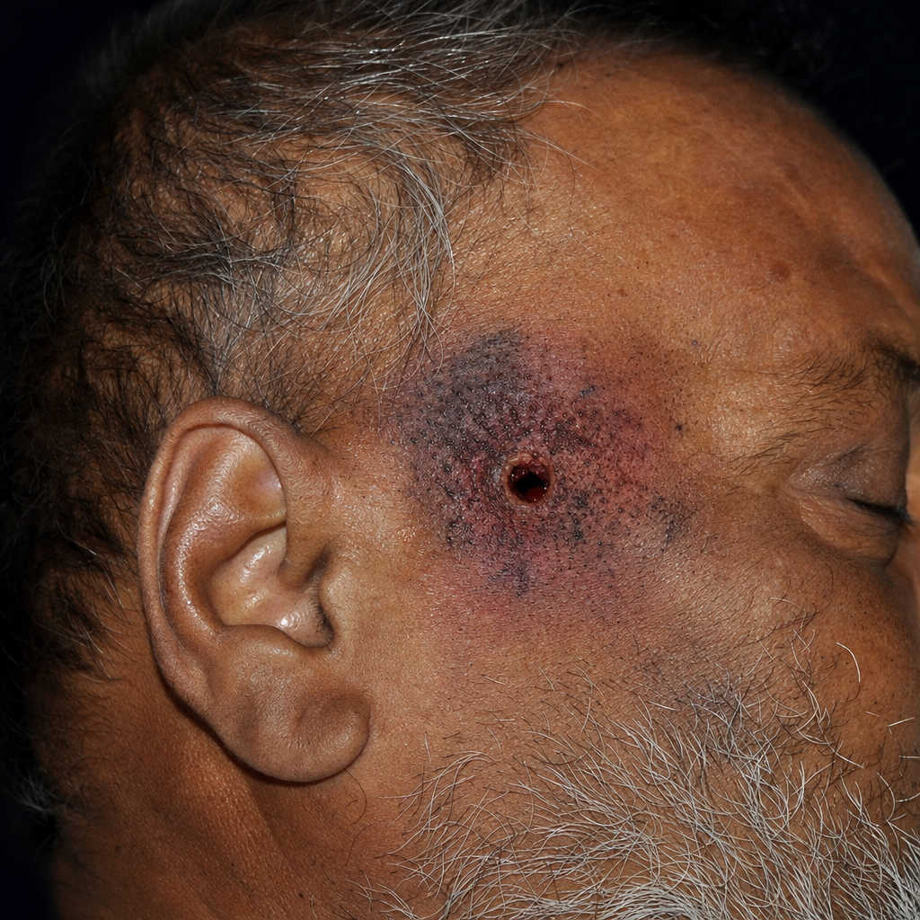
70-year-old man who died suddenly has the finding shown here on external examination at autopsy. Which of the following is the most likely diagnosis?
The main danger with low tension alternating current is
If a fracture gives the pattern of the striking surface of the weapon it is called
You are conducting an autopsy on a patient and you find ladder tears near the main lesion on a major blood vessel. What is the most likely cause of the injury and subsequent death of the person?
Practice by Chapter
Mechanical Injuries
Practice Questions
Transportation Injuries
Practice Questions
Fall from Height
Practice Questions
Blunt Force Trauma
Practice Questions
Sharp Force Trauma
Practice Questions
Ballistic Injuries
Practice Questions
Burn Injuries
Practice Questions
Drowning
Practice Questions
Electrocution
Practice Questions
Lightning Injuries
Practice Questions
Explosion Injuries
Practice Questions
Pattern Injuries and Their Recognition
Practice Questions
Want unlimited practice?
Get full access to all questions, explanations, and performance tracking.
Scan to download app