
Injuries and Their Significance — MCQs

On this page
Ante-mortem blisters differ from post-mortem blisters by which of the following characteristics?
In bursting of the skull due to firearm injury, a large portion of the brain may be thrown out of the bursting skull and found relatively intact. What is this phenomenon called?
What determines the destructive power of a bullet?
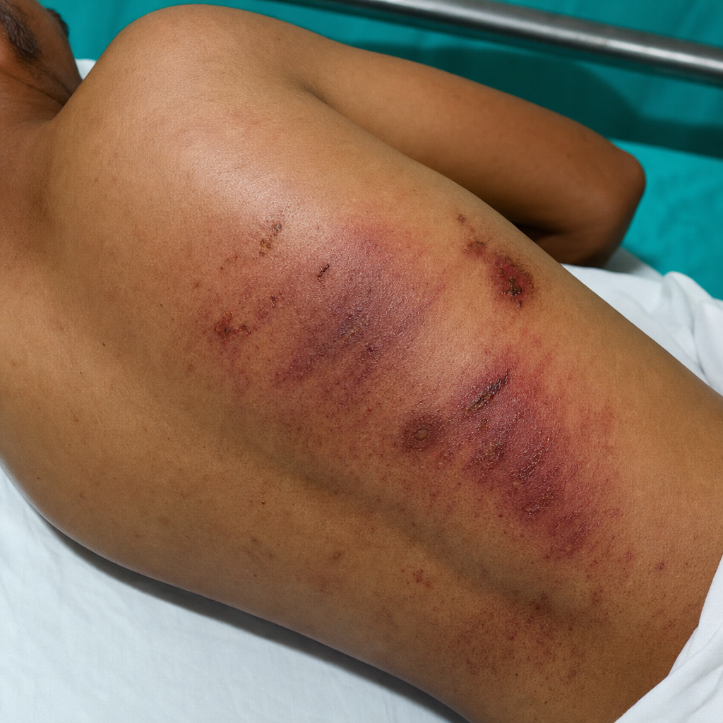
The type of injury is:
Ante-mortem burns differ from post-mortem burns by all of the following findings, EXCEPT:
What is the 'Di collar' seen in?
Unaker's fracture is seen at the level of which cervical vertebra?
The commonest type of abrasion seen in road traffic accidents is:
What type of abrasions are present on the dead body?
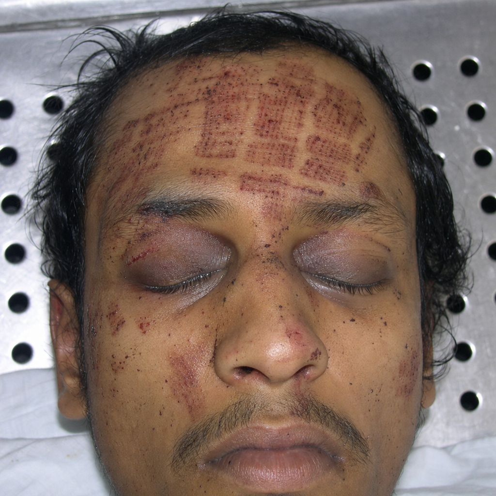
Blackening of the eye after a forehead injury is typically due to which of the following phenomena?
Practice by Chapter
Mechanical Injuries
Practice Questions
Transportation Injuries
Practice Questions
Fall from Height
Practice Questions
Blunt Force Trauma
Practice Questions
Sharp Force Trauma
Practice Questions
Ballistic Injuries
Practice Questions
Burn Injuries
Practice Questions
Drowning
Practice Questions
Electrocution
Practice Questions
Lightning Injuries
Practice Questions
Explosion Injuries
Practice Questions
Pattern Injuries and Their Recognition
Practice Questions
Want unlimited practice?
Get full access to all questions, explanations, and performance tracking.
Scan to download app