
Injuries and Their Significance — MCQs

On this page
Waddell's trial does not include which of the following injuries?
What does the tail of a wound indicate?
Which of the following is not altered by rifling a gun?
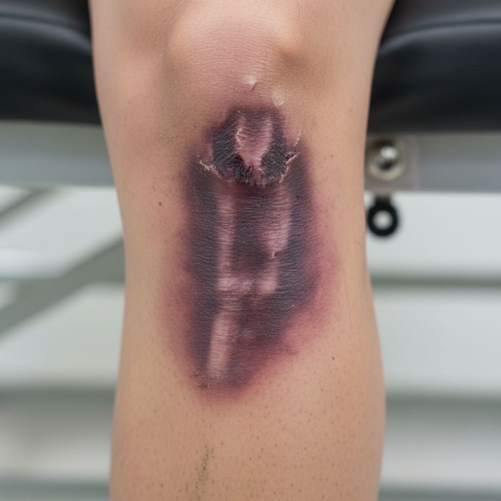
The following injury is:
Which of the following skull fractures is called a Motorcyclist's fracture?
More than 5% carboxyhemoglobin is indicative of what condition?
A 25-year-old female was found with 100% burns on her body. The tongue was protruding, the body was in a pugilistic attitude with heat ruptures, peeling of the skin, and heat hematoma and heat fractures of the skull. Carboxyhemoglobin was 25% and soot particles were present in the trachea. Which combination of two findings will establish that the burns were ante-mortem in nature?
Black gunpowder contains all of the following, except?
Which of the following areas, when bruised, does NOT show typical color changes?
Which of the following markings is NOT typically seen on a fired cartridge case?
Practice by Chapter
Mechanical Injuries
Practice Questions
Transportation Injuries
Practice Questions
Fall from Height
Practice Questions
Blunt Force Trauma
Practice Questions
Sharp Force Trauma
Practice Questions
Ballistic Injuries
Practice Questions
Burn Injuries
Practice Questions
Drowning
Practice Questions
Electrocution
Practice Questions
Lightning Injuries
Practice Questions
Explosion Injuries
Practice Questions
Pattern Injuries and Their Recognition
Practice Questions
Want unlimited practice?
Get full access to all questions, explanations, and performance tracking.
Scan to download app