
Otology — MCQs

On this page
Prussak's space is situated in which part of the middle ear?
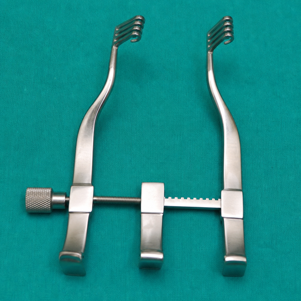
All of the following are uses of the given instrument except:
Which part of the middle ear is most commonly involved in otosclerosis?
A child presents with a three-month history of left ear hearing loss. On examination, foul-smelling purulent discharge is observed, along with a perforation in the pars flaccida. What is the most appropriate management?
Paracusis willis is a feature of which condition?
Fisch classification is used for which type of tumor?
What procedure involves widening of the cartilaginous part of the external auditory canal?
Pulsatile tinnitus in the ear is most commonly due to which of the following conditions?
Galle's test is used for which of the following conditions?
What is the most mobile part of the tympanic membrane?
Practice by Chapter
Tympanic Membrane Perforation
Practice Questions
Cholesteatoma
Practice Questions
Tympanoplasty Techniques
Practice Questions
Ossicular Chain Reconstruction
Practice Questions
Mastoidectomy
Practice Questions
Stapedectomy
Practice Questions
Implantable Hearing Devices
Practice Questions
Congenital Aural Atresia
Practice Questions
Otologic Trauma
Practice Questions
Glomus Tumors
Practice Questions
Facial Nerve Decompression
Practice Questions
Rehabilitative Audiology
Practice Questions
Want unlimited practice?
Get full access to all questions, explanations, and performance tracking.
Scan to download app