Which one of the following conditions produces sensorineural deafness ?
A female patient's pure tone audiometry (PTA) findings show the presence of a Carhart's notch. Which of the following specific clinical signs can be seen in this patient?
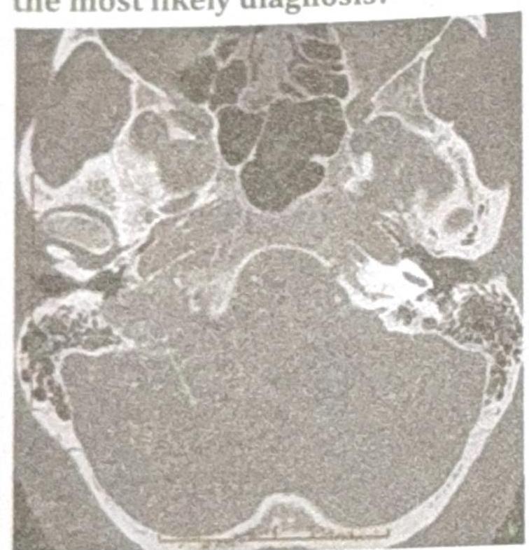
A patient presents with conductive hearing loss, pulsatile tinnitus, and a positive Phelps sign. Based on the CT scan image provided, what is the most likely diagnosis?

Gelle's test is done in?
Positive Rinne test is seen in -
Most difficult site to remove cholesteatoma from the sinus tympani is related to:
Columellar tympanoplasty is -
All of the following techniques are used to control bleeding from bone during mastoid surgery except:
Picket fence fever is a feature of -
Iatrogenic traumatic facial nerve palsy is MOST commonly produced during which of the following surgical procedures?
Explanation: ***Mixed otosclerosis*** - **Otosclerosis** is a bone remodeling disease primarily affecting the **ossicles** and **otic capsule**. - **Mixed otosclerosis** involves both **stapes fixation (conductive component)** and **cochlear involvement (sensorineural component)**. - Among the given options, this is the **only condition that produces sensorineural hearing loss**, although it presents as a mixed hearing loss with both conductive and sensorineural components. - The **cochlear otosclerosis** component causes the sensorineural deafness through involvement of the **otic capsule** and **cochlear structures**. *Atelectatic middle ear* - This condition involves the collapse of the **tympanic membrane** onto the ossicles due to **negative middle ear pressure**. - It causes **pure conductive hearing loss** by impairing sound transmission through the middle ear, with no sensorineural component. *Eustachian tube blockage* - Blockage of the **Eustachian tube** leads to **negative pressure** in the middle ear space. - This results in **pure conductive hearing loss** due to impaired movement of the tympanic membrane and ossicles, with no sensorineural component. *Tympanic membrane rupture* - A rupture in the **tympanic membrane** creates a direct opening between the external ear canal and the middle ear. - This condition causes **pure conductive hearing loss** by disrupting the normal sound conduction mechanism, with no sensorineural component.
Explanation: ***Schwartz sign*** - A **Carhart's notch** (bone conduction dip at 2000 Hz) is a characteristic finding in **otosclerosis**, a condition where the stapes bone becomes fixed. - The **Schwartz sign** (reddish/pinkish hue behind the tympanic membrane) indicates **active otosclerosis** with increased vascularity in the promontory. - This is a **specific clinical sign** directly associated with otosclerosis and would be the expected finding on otoscopy. *Hitselberger sign* - The **Hitselberger sign** refers to **hypesthesia** (numbness or decreased sensation) in the **posterior external auditory canal wall**. - It is associated with **acoustic neuromas** (vestibular schwannomas) due to compression of the facial nerve, not otosclerosis. *Hennebert sign* - The **Hennebert sign** involves **nystagmus or vertigo** induced by changes in external ear canal pressure (fistula test). - It is typically seen in conditions causing a **perilymphatic fistula**, such as **Meniere's disease** or **syphilitic labyrinthitis**, not otosclerosis. *Rinne test negative (conductive hearing loss)* - While otosclerosis does cause **conductive hearing loss** with a negative Rinne test (bone conduction > air conduction), this is a **general audiometric finding**, not a specific clinical sign. - A negative Rinne test can occur in **any cause of conductive hearing loss** (chronic otitis media, ossicular discontinuity, cholesteatoma, etc.). - The question asks for a **specific clinical sign**, making the **Schwartz sign** the most appropriate answer as it specifically indicates otosclerosis.
Explanation: ***Glomus Jugulare*** - The symptoms of **conductive hearing loss**, **pulsatile tinnitus**, and a **positive Phelps sign** (destruction of the bone separating the carotid artery from the jugular bulb on CT) are classic for a glomus jugulare tumor. - The CT scan image, showing a destructive lesion in the **jugular foramen** region, further supports this diagnosis, as these tumors originate from the paraganglia of the jugular bulb. *Glomus Tympanicum* - While it can cause conductive hearing loss and pulsatile tinnitus, a glomus tympanicum tumor is typically **confined to the middle ear cavity** and does not involve bone erosion of the jugular foramen as indicated by a positive Phelps sign. - The CT findings would show a mass in the middle ear, often behind an intact tympanic membrane, rather than extensive bone destruction in the jugular region. *Acoustic Neuroma* - This tumor, also known as a **vestibular schwannoma**, primarily causes **sensorineural hearing loss**, **tinnitus** (often non-pulsatile), and **balance issues**, not conductive hearing loss or pulsatile tinnitus. - An acoustic neuroma is located in the **cerebellopontine angle** and internal auditory canal, and the CT scan would show a mass in this specific location, not necessarily bone erosion of the jugular foramen. *Cholesteatoma* - A cholesteatoma is a **destructive, expanding growth of keratinizing squamous epithelium** in the middle ear or mastoid, typically presenting with **conductive hearing loss**, **otorrhea**, and sometimes headaches. - It does not typically cause pulsatile tinnitus unless it's very large and vascularized, nor does it present with the characteristic destruction of the jugular foramen seen with a positive Phelps sign.
Explanation: ***Otosclerosis*** - **Gelle's test** is primarily used to diagnose **otosclerosis**, a condition causing **conductive hearing loss** due to abnormal bone growth in the middle ear. - The test assesses changes in **bone conduction hearing** in response to alterations in external ear canal pressure, which is characteristic of a fixed stapes footplate in otosclerosis. *Traumatic deafness* - **Traumatic deafness** can result from direct injury to the ear, but **Gelle's test** is not a primary diagnostic tool for this type of hearing loss. - Diagnosis typically involves evaluating the **nature and extent of the trauma** and other audiometric tests. *Senile deafness* - Also known as **presbycusis**, **senile deafness** is a **sensorineural hearing loss** associated with aging. - **Gelle's test** assesses changes in bone conduction with pressure, making it less relevant for diagnosing age-related nerve damage. *Serous otitis media* - **Serous otitis media** involves fluid accumulation in the middle ear, leading to **conductive hearing loss**. - While it causes conductive hearing loss, **Gelle's test** is not the definitive diagnostic test; **tympanometry** and **audiometry** are more commonly used.
Explanation: ***Presbycusis*** - A **positive Rinne test** indicates that **air conduction is better than bone conduction (AC > BC)**, which is normal. - In presbycusis, which is a **sensorineural hearing loss**, the inner ear is affected. While hearing is impaired, the *relationship between air and bone conduction remains normal*, thus a positive Rinne test is maintained. *Wax impacted ear* - This causes a **conductive hearing loss** by obstructing the ear canal. - In conductive hearing loss, **bone conduction is better than air conduction (BC > AC)**, resulting in a **negative Rinne test**. *CSOM* - **Chronic Suppurative Otitis Media (CSOM)** involves infection and perforation of the tympanic membrane and/or damage to ossicles. - This leads to **conductive hearing loss**, where **bone conduction is better than air conduction**, resulting in a **negative Rinne test**. *Otosclerosis* - This condition involves **abnormal bone growth** in the middle ear, particularly around the stapes footplate, leading to fixation. - It causes a **conductive hearing loss**, characterized by **bone conduction being better than air conduction**, meaning a **negative Rinne test**.
Explanation: **Posterior facial ridge** - The **posterior facial ridge** forms a critical anatomical barrier, often **obscuring the sinus tympani** and making cholesteatoma removal difficult due to limited access and visibility. - Its presence necessitates careful surgical technique to avoid **facial nerve injury** while ensuring complete eradication of disease. *Anterior facial ridge* - The **anterior facial ridge** is less directly involved in defining the access to the sinus tympani, thus not posing as significant a surgical challenge for cholesteatoma removal in this specific area. - Its anatomical position typically allows for better visualization and maneuverability compared to the posterior ridge. *Epitympanum* - The **epitympanum**, while a common site for cholesteatoma, is generally more accessible surgically than the sinus tympani because of its location superior to the tympanic membrane. - Its removal often involves mastoidectomy or atticotomy, which provides direct views. *Hypotympanum* - The **hypotympanum** is located inferior to the tympanic membrane and is typically more open and accessible, allowing for easier visualization and removal of cholesteatoma. - It does not present the same deep, narrow, and obstructed surgical field as the sinus tympani, especially in relation to the facial nerve.
Explanation: ***Type IV*** - **Columellar tympanoplasty** is characterized by the placement of the graft directly onto the **stapes footplate**, creating a **columella effect** to transmit sound. - This type of tympanoplasty is performed when the entire **ossicular chain** is missing, except for a mobile stapes footplate, effectively bypassing the malleoincudal complex. *Type III* - Involves placing the graft directly onto the **head of the stapes**, requiring a mobile stapes with an intact superstructure. - This method is used when the **malleus and incus are missing**, but the stapes is functional. *Type I* - Also known as **myringoplasty**, this type involves repairing a **perforated tympanic membrane** when the ossicular chain is intact and functional. - The goal is to restore the integrity of the eardrum to improve hearing by closing the perforation. *Type II* - This involves repairing a perforated tympanic membrane while also addressing minor **ossicular chain defects**. - The graft is placed on the **incus** (or malleus) after reconstructing a portion of the ossicular chain, often due to erosion of the incus.
Explanation: ***Cutting drill over the bleeding area*** - A **cutting drill** is designed to remove bone rapidly and would *exacerbate bleeding* from bone by causing further trauma to blood vessels, not control it. - Its sharp flutes create a cutting action that is unsuitable for managing hemorrhage, unlike instruments designed for coagulation or surface abrasion. *Diamond drill over the bleeding area* - A **diamond drill** is effective for controlling superficial bone bleeding because its abrasive action polishes and **occludes small vascular channels**, creating less trauma than a cutting drill. - The friction generated by the diamond burr can also contribute to **thermal coagulation** of small vessels. *Bipolar cautery over the bleeding area* - **Bipolar cautery** uses electrical current to **coagulate bleeding vessels** directly in the bone, effectively sealing them. - It is a common and effective method for controlling bone bleeding, especially for larger vascular channels exposed during drilling. *Bone wax* - **Bone wax** (e.g., paraffin wax or beeswax) is a sterile mixture used to mechanically **occlude bleeding capillaries and small vessels** within the cut edges of bone. - It acts as a **physical barrier** to bleeding, providing immediate hemostasis when pressed onto the bleeding bone surface.
Explanation: ***Lateral sinus thrombophlebitis*** - **Picket fence fever**, characterized by high spiking fevers followed by abrupt drops to normal or subnormal temperature, is a classic symptom of **lateral sinus thrombophlebitis** due to septic emboli. - This condition is a serious complication of otitis media or mastoiditis, where infection from the middle ear or mastoid spreads to the **dural venous sinuses**. *Acute mastoiditis* - Acute mastoiditis typically presents with **postauricular pain**, swelling, and erythema, often accompanied by ear discharge and fever, but not specifically picket fence fever. - While mastoiditis can lead to lateral sinus thrombophlebitis, it is the underlying infection, not the defining feature of the fever pattern. *Atticoantral CSOM* - **Atticoantral chronic suppurative otitis media (CSOM)**, also known as cholesteatoma, is characterized by chronic ear discharge, hearing loss, and often cholesteatoma formation. - While it can lead to complications, significant systemic fever patterns like picket fence fever are not typical for uncomplicated atticoantral CSOM itself. *Bell's Palsy* - **Bell's Palsy** is an acute peripheral facial nerve paralysis of unknown cause, presenting with unilateral facial weakness or paralysis. - It is not associated with fever, particularly not the cyclical spiking pattern described as picket fence fever, as it is a neurological condition.
Explanation: ***Mastoidectomy*** - **Mastoidectomy** involves extensive bone removal around the **facial nerve's mastoid segment**, making it the most vulnerable during this procedure due to its complex anatomical course. - The surgery's depth and proximity to the fallopian canal increase the risk of accidental nerve injury from drilling or instrumentation. *Ossiculoplasty* - **Ossiculoplasty** primarily involves repairing or replacing the **ossicles** in the middle ear, generally not requiring manipulation close to the facial nerve. - While the nerve is in the vicinity, direct trauma is rare compared to extensive bone work. *Myringoplasty* - **Myringoplasty** is a relatively superficial procedure involving the repair of the **tympanic membrane**, far from the main course of the facial nerve. - The surgical field is typically limited to the external auditory canal and middle ear cavity, posing minimal risk to the nerve. *Stapedectomy* - **Stapedectomy** involves operating on the **stapes bone** in the oval window, which is near the horizontal segment of the facial nerve, but less frequently injured compared to mastoid surgery. - Although the nerve is identified and protected, the extent of bone removal and proximity is less than in a full mastoidectomy.
Tympanic Membrane Perforation
Practice Questions
Cholesteatoma
Practice Questions
Tympanoplasty Techniques
Practice Questions
Ossicular Chain Reconstruction
Practice Questions
Mastoidectomy
Practice Questions
Stapedectomy
Practice Questions
Implantable Hearing Devices
Practice Questions
Congenital Aural Atresia
Practice Questions
Otologic Trauma
Practice Questions
Glomus Tumors
Practice Questions
Facial Nerve Decompression
Practice Questions
Rehabilitative Audiology
Practice Questions
Get full access to all questions, explanations, and performance tracking.
Start For Free