Identify the treatment option with the image given below:
A 35-year-old woman presented to the clinic with the symptoms of hearing loss and pulsatile tinnitus. Further examination reveals conductive hearing loss with the Rinne test negative. A reddish mass is seen behind the tympanic membrane. What is the most likely diagnosis for this patient?
A patient presents to the clinic with a history of chronic ear drainage and hearing loss for several months. Otoscopic examination reveals a central perforation of the tympanic membrane. Pure tone audiometry confirms a conductive hearing loss. Based on these findings, what is the most appropriate next step in the definitive management of this patient's condition?
A 30-year-old male presents to the clinic with a history of recurrent ear infections and a recent "popping" sensation in his left ear. Otoscopic examination of the left ear reveals a central perforation of the tympanic membrane. The pure tone audiometry (PTA) results show the following: Right Ear: Air conduction and bone conduction thresholds are within normal limits. Left Ear: A significant air-bone gap is present, with bone conduction thresholds within the normal range. Based on these findings, what is the most likely diagnosis regarding the type of hearing loss?
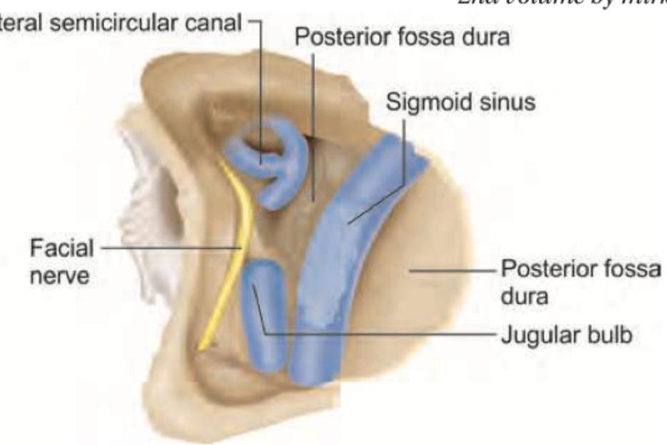
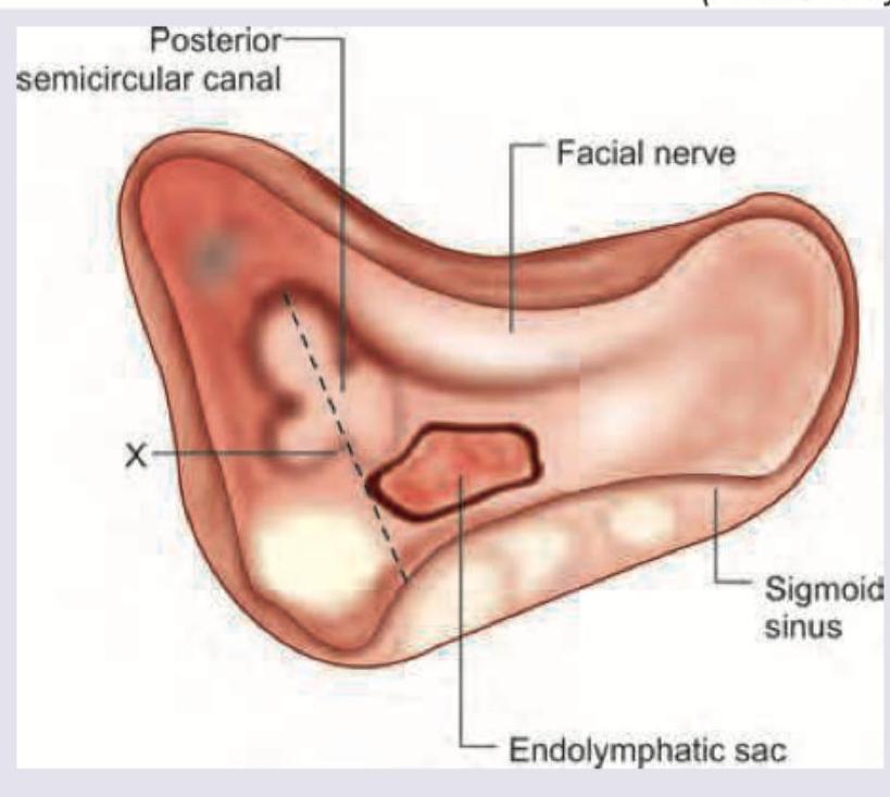
Identify the structure marked in the given image:

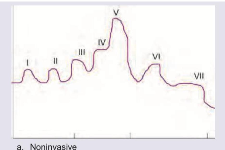
All are true about the recording shown in the image except:

Which of the following is a contraindication to the procedure shown below?

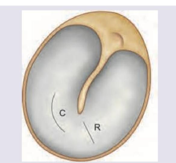
The tympanogram shown below is seen in which of the following conditions?

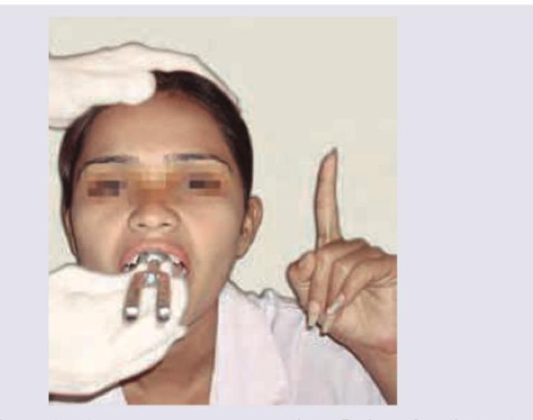
Which test is being performed in the patient?

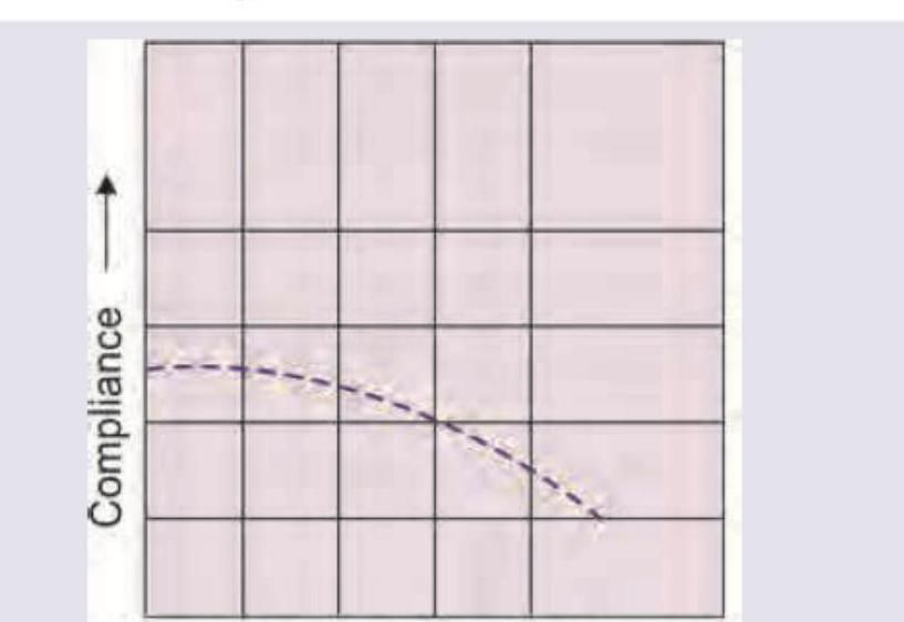
Identify the line shown in the given image:

Explanation: ***Type 3 tympanoplasty*** - This procedure, also known as **myringostapediopexy**, involves placing a graft directly onto the head of the stapes, as depicted in the image. - It is indicated when both the **malleus** and **incus** are eroded or absent, but the stapes is intact and mobile, allowing for sound transmission directly to the stapes. *Type 1 Tympanoplasty* - This is a simple repair of a tympanic membrane perforation, also called **myringoplasty**, where the graft is placed to close the hole. - This procedure is only suitable when the **ossicular chain** is completely intact and mobile, which is not the case shown. *Type 2 tympanoplasty* - This is performed when the **malleus** is eroded, and the graft is placed onto the **incus** or the remaining part of the malleus. - The image shows that the incus is also absent, as the graft is placed directly on the stapes, making this option incorrect. *Type 4 tympanoplasty* - This procedure is indicated when only the **stapes footplate** remains and is mobile; the graft is placed directly on the footplate. - This creates a very shallow middle ear space, essentially shielding only the round window and Eustachian tube opening, a more extensive reconstruction than shown.
Explanation: ***Glomus tumour (Correct Answer)*** - **Pulsatile tinnitus** combined with **conductive hearing loss** and a **reddish retrotympanic mass** forms the classic diagnostic triad for glomus tympanicum (a paraganglioma arising from glomus bodies). - The **reddish vascular mass** behind the tympanic membrane is pathognomonic, sometimes called the **'rising sun sign'** or demonstrating **Brown's sign** (blanching with pneumatic otoscopy). - This slow-growing, highly vascular tumor characteristically presents with these features in middle-aged adults. *Acute otitis media (Incorrect)* - This acute bacterial infection presents with rapid onset of **otalgia** (ear pain), **fever**, and a bulging, intensely red tympanic membrane. - While it causes temporary conductive hearing loss due to **purulent fluid** accumulation, it lacks the chronic presentation and **pulsatile tinnitus** characteristic of vascular masses. - The clinical course is acute (days), not chronic like glomus tumors. *Chronic otitis media (Incorrect)* - Defined by chronic inflammation typically resulting in **tympanic membrane perforation**, recurrent **otorrhea** (ear discharge), and possible **cholesteatoma** formation. - While it causes conductive hearing loss, it does **not** produce **pulsatile tinnitus** or a **reddish, vascular retrotympanic mass**. - The tympanic membrane shows perforation or scarring, not an intact membrane with a vascular mass behind it. *Serous otitis media (Incorrect)* - Also known as **otitis media with effusion**, involves sterile, non-purulent fluid in the middle ear from **eustachian tube dysfunction**. - The tympanic membrane appears dull, retracted, or shows **air-fluid levels** and bubbles, but does not show a vascular mass. - This condition does **not** cause **pulsatile tinnitus** and the fluid is serous, not vascular tissue.
Explanation: ***Myringoplasty*** - This procedure involves the surgical repair of the **tympanic membrane (TM)** perforation (a central perforation in this case) and is the definitive treatment for chronic otitis media, tubotympanic type (safe CSOM). - Repairing the TM perforation eliminates the source of chronic drainage and optimally helps in resolving the **conductive hearing loss** by restoring the vibrating surface. *Modified radical mastoidectomy* - This procedure is primarily reserved for **unsafe chronic otitis media** (Atticoantral disease), typically involving **cholesteatoma** or extensive irreversible mucosal disease in the mastoid. - Since the patient has a **central perforation** (safe type) without mention of retraction pockets or cholesteatoma, this extensive surgery is overmanagement and not indicated. *Exploratory tympanostomy* - This term generally refers to making an incision in the TM (myringotomy) or placing a **ventilation tube (grommet)**, typically done for otitis media with effusion (serous otitis media). - It does not address the underlying **chronic irreversible perforation** of the TM causing the current symptoms; surgical repair is required. *Immediate commencement of broad-spectrum oral antibiotics* - Antibiotics are useful for managing acute exacerbations of CSOM (controlling active discharge or infection) but are not the **definitive treatment** for the underlying structural pathology (chronic perforation). - They cannot **repair the tympanic membrane** or permanently restore hearing; the structural defect requires surgical intervention (myringoplasty).
Explanation: ***Left conductive hearing loss*** - A conductive hearing loss is defined on PTA by the presence of a significant **air-bone gap** (AC threshold worse than BC threshold) with **normal bone conduction** thresholds. - The otoscopic finding of a **central perforation of the tympanic membrane** indicates a pathology in the middle ear/conducting mechanism, which is the classical cause of a conductive loss. *Left sensorineural hearing loss* - This type of loss occurs when both **air conduction** and **bone conduction** thresholds are equally reduced (no air-bone gap) and are outside the normal limits. - It indicates a problem with the **cochlea** or the **auditory nerve**, which contradicts the normal bone conduction found in the left ear. *Right conductive hearing loss* - The PTA results for the right ear showed **air and bone conduction thresholds** within normal limits, confirming **normal hearing** in the right ear. - Therefore, a hearing loss of any type in the right ear is ruled out by the objective audiometric data. *Right sensorineural hearing loss* - This would require both air and bone conduction thresholds in the right ear to be significantly elevated (impaired), which is inconsistent with the **normal audiometry findings** reported for the right ear.
Explanation: ***Lateral SCC*** - The image provided clearly labels the "Lateral semicircular canal" with a line pointing to the structure in question, confirming its identity. - The **semicircular canals** are part of the bony labyrinth of the inner ear, crucial for maintaining **balance**. *Facial nerve* - The **facial nerve** is also labeled in the diagram but points to a thin, yellow structure separate from the blue curved structure. - The facial nerve's primary role is controlling **facial expressions** and taste sensation. *Sino-dural angle* - The **sino-dural angle** refers to the junction of the sigmoid sinus and the posterior fossa dura, which is not specifically highlighted or pointed to as the structure in question. - This anatomical landmark is important in **otologic surgery** as a reference point. *Sigmoid sinus* - The **sigmoid sinus** is a venous channel labeled on the right side of the diagram, appearing as a larger blue structure. - It collects blood from the brain and drains into the **internal jugular vein**.
Explanation: ***Wave I originates from proximal part of Cranial nerve VIII*** - **Wave I** of the ABR actually originates from the **distal portion of cranial nerve VIII**, specifically near the cochlea. - The more proximal parts of the auditory nerve and brainstem generate subsequent waves (II-VII). *Noninvasive* - Auditory brainstem response (**ABR**) testing is indeed a **noninvasive** procedure. - It involves placing electrodes on the scalp to record electrical activity in response to auditory stimuli. *Test integrity of central auditory pathways* - ABR measures electrical activity as sound travels from the cochlea through the **auditory nerve** and into the **brainstem**. - This makes it an excellent tool for assessing the **integrity of the central auditory pathways**, identifying neurological issues. *Measures hearing in range of 1000-4000 Hz* - ABR testing primarily evaluates hearing sensitivity in the **higher frequency range**, typically 1000-4000 Hz. - This range is crucial for speech understanding and is often the first to be affected in certain types of hearing loss.
Explanation: ***Glomus tumor*** - A **glomus tumor** as a contraindication because it is a highly vascular tumor in the middle ear, and performing a myringotomy (as depicted by the incisions 'C' and 'R' on the tympanic membrane) could lead to **severe hemorrhage**. - Identification of this tumor, often presenting as a **pulsatile red mass** behind the tympanic membrane, mandates further evaluation and planned surgical excision rather than a simple myringotomy. *Osteosclerosis* - **Osteosclerosis** refers to increased bone density and is not directly related to the tympanic membrane itself. - It would not typically contraindicate a myringotomy, which is a procedure involving an incision in the eardrum. *Myringosclerosis* - **Myringosclerosis** involves **calcified plaques** on the tympanic membrane, which indicates a thickened, scarred, and often less mobile eardrum. - While it may make the membrane slightly more rigid, it does not contraindicate a myringotomy when indicated for conditions like otitis media with effusion. *Thick tympanic membrane* - A **thick tympanic membrane** is often a result of chronic inflammation or past infections. - While it might require a sharper or stronger incision, it is not a direct contraindication to myringotomy, as the procedure is still feasible and can be necessary for drainage or ventilation.
Explanation: ***Middle ear fluid*** - The tympanogram shown is a **Type B** tympanogram, which is characteristic of **middle ear fluid**. - A Type B tympanogram indicates **severely reduced or no compliance** of the tympanic membrane, suggesting the middle ear is filled with fluid and cannot vibrate normally. *Otosclerosis* - Otosclerosis typically causes a **Type As** tympanogram, characterized by **reduced compliance but normal middle ear pressure**. - This is due to the **stiffening of the ossicular chain**, primarily the stapes footplate. *Ossicular disruption* - Ossicular disruption typically results in a **Type Ad** tympanogram, showing **excessively high compliance** (a deep, exaggerated peak). - This occurs because the ossicular chain is discontinuous, leading to reduced impedance and increased movement of the tympanic membrane. *Eustachian tube dysfunction* - Eustachian tube dysfunction often presents as a **Type C** tympanogram, characterized by a **negative middle ear pressure** with normal or near-normal compliance. - This negative pressure results from the Eustachian tube's inability to ventilate the middle ear, leading to absorption of air and retraction of the eardrum.
Explanation: ***Weber test*** - The image depicts a **tuning fork placed on the patient's central incisors**, which is a technique used to perform the Weber test. - The Weber test assesses **bone conduction** and helps differentiate between conductive and sensorineural hearing loss by determining if the sound lateralizes to one ear. *Rinne test* - The Rinne test compares **air conduction to bone conduction** in the same ear. - It involves placing a vibrating tuning fork on the **mastoid process** and then holding it in front of the ear canal. *Schwabach test* - The Schwabach test compares the patient's **bone conduction** with that of the examiner, assuming the examiner has normal hearing. - It involves placing a vibrating tuning fork on the **mastoid process** of both the patient and examiner. *Absolute bone conduction test* - This test is a variant of the Schwabach test where the **external auditory canal is occluded** to eliminate air conduction. - It aims to assess bone conduction exclusively, but the method shown in the image (tuning fork on incisors for lateralization) is characteristic of the Weber test.
Explanation: ***Donaldson line*** - The image illustrates the posterior aspect of the petrous temporal bone, showing the relationship between the **facial nerve**, **posterior semicircular canal**, and the **endolymphatic sac**. - The dashed line directly passes superomedial to the **endolymphatic sac** and inferolateral to the **posterior semicircular canal**, which is characteristic of Donaldson's line, used as a surgical landmark. *Ohngren's line* - Ohngren's line is a radiological landmark used in the context of **maxillary sinus tumors**. - It runs from the **medial canthus of the eye** to the **angle of the mandible**, dividing the maxillary sinus. *Weber Ferguson line* - The Weber-Ferguson incision is a **facial incision** used for access to the **maxillary sinuses and midface**. - It is a surgical approach, not an anatomical line delineating internal structures. *Frankfurt line* - The Frankfurt horizontal plane, or Frankfurt line, is an **anthropometric landmark** used in craniometry and orthodontics. - It is defined by the superior border of the **external auditory meatus** and the inferior orbital rim.
Tympanic Membrane Perforation
Practice Questions
Cholesteatoma
Practice Questions
Tympanoplasty Techniques
Practice Questions
Ossicular Chain Reconstruction
Practice Questions
Mastoidectomy
Practice Questions
Stapedectomy
Practice Questions
Implantable Hearing Devices
Practice Questions
Congenital Aural Atresia
Practice Questions
Otologic Trauma
Practice Questions
Glomus Tumors
Practice Questions
Facial Nerve Decompression
Practice Questions
Rehabilitative Audiology
Practice Questions
Get full access to all questions, explanations, and performance tracking.
Start For Free