
Diseases of the Oral Cavity and Salivary Glands — MCQs

On this page
What is a constant feature associated with a radicular cyst?
What is the most common site of sialolithiasis?
Which of the following is untrue regarding Ludwig's angina?
Multiple painful ulcers on the tongue are seen in all the following conditions EXCEPT?
A patient presents with bilateral diffuse swelling in the neck below the chin. Intraorally, the lower left molar is infected. Severe cellulitis in the submandibular, sublingual, and submental spaces may cause death due to which of the following?
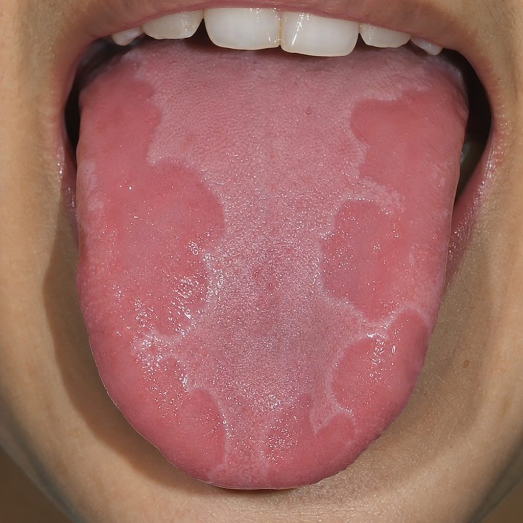
White raised painless areas on peeling exhibit painful erythematous areas in case of?
Which of the following statements regarding hard palate tumors is true?
Which of the following statements regarding tumors of the hard palate is false?
Reverse smoking is a known risk factor for which of the following conditions?
Which of the following is the diagnosis of the condition shown?
Practice by Chapter
Stomatitis
Practice Questions
Oral Ulcers
Practice Questions
Oral Leukoplakia
Practice Questions
Oral Cancers
Practice Questions
Sialadenitis
Practice Questions
Sialolithiasis
Practice Questions
Salivary Gland Tumors
Practice Questions
Ranula
Practice Questions
Xerostomia
Practice Questions
Sjögren's Syndrome
Practice Questions
Oral Manifestations of Systemic Diseases
Practice Questions
Temporomandibular Joint Disorders
Practice Questions
Want unlimited practice?
Get full access to all questions, explanations, and performance tracking.
Scan to download app