Acute non-suppurative sialadenitis is most commonly seen in which condition?
What is Epulis?
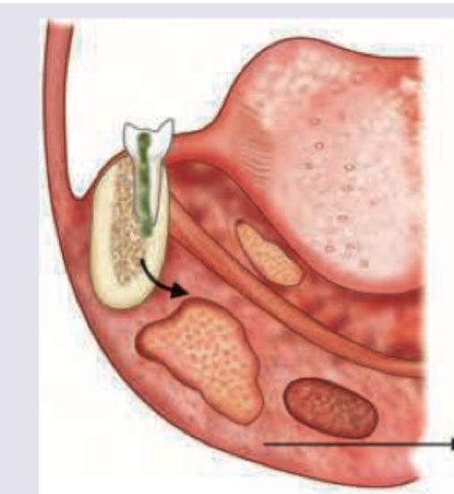
During the extraction of an upper first molar, the mesiobuccal root is missing and suspected to have been pushed into the maxillary sinus. What is the recommended method to determine antral perforation?
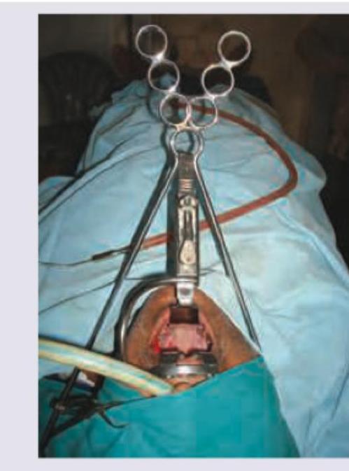
Which is incorrect about the instrument shown?

A patient with dental caries has developed a swelling in the area shown in the image. Diagnosis is:

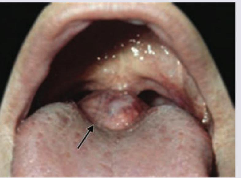
A 16-year-old patient complains of difficulty in swallowing, difficulty in talking and sometimes difficulty in breathing. On physical examination the presentation is similar to that shown in the picture. What would be the probable diagnosis?

A 6-year-old boy has recurrent history of pain and swelling below his left ear, which generally lasts for 3–7 days and improves mildly after a course of antibiotics. Sialography shows punctate sialectasis. He should be treated by
What is a clinical feature of mouth breathing?
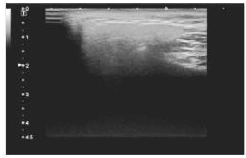
A 45-year-old patient complained of pain on one side of the neck. She is afraid of eating food as it worsens the pain. An ultrasound of the salivary glands is shown below. What is the most likely diagnosis?

Which anatomical location is often referred to as the 'graveyard of the ENT surgeon' due to its association with undetected malignancies?
Explanation: **Explanation:** **Mumps** is the most common cause of **acute non-suppurative sialadenitis**. It is a viral infection caused by the *Paramyxovirus*. Unlike bacterial infections, viral sialadenitis typically presents with sudden, painful swelling of the salivary glands (most commonly the parotid) without the formation of pus (non-suppurative). The inflammation is interstitial, and the saliva remains clear, though the amylase levels may be elevated. **Analysis of Incorrect Options:** * **A. Acute bacterial sialadenitis:** This is a **suppurative** condition, usually caused by *Staphylococcus aureus*. It is characterized by exquisite pain, fever, and the expression of frank pus from the duct (Stensen’s duct) upon massage. * **C. Chronic bacterial sialadenitis:** This refers to recurrent or long-standing inflammation, often secondary to ductal obstruction (sialolithiasis). It involves progressive destruction of the acini and fibrosis rather than an acute non-suppurative phase. * **D. Necrotizing sialometaplasia:** This is a benign, self-limiting inflammatory condition typically affecting the minor salivary glands of the hard palate. It mimics malignancy clinically and histologically but is not a primary cause of acute generalized sialadenitis. **High-Yield Clinical Pearls for NEET-PG:** * **Mumps:** Most common complication in children is **meningoencephalitis**; in post-pubertal males, it is **orchitis** (usually unilateral). * **Diagnosis:** Primarily clinical; however, an elevated **Serum Amylase** is a classic biochemical marker. * **Bacterial Sialadenitis:** Often seen in dehydrated, elderly, or postoperative patients ("Surgical Parotitis"). * **Treatment for Mumps:** Purely symptomatic (hydration and analgesics); antibiotics have no role unless secondary bacterial infection occurs.
Explanation: **Explanation:** **Epulis** is a non-specific clinical term used to describe any tumor-like swelling or growth located on the **gingiva** (gums) or alveolar mucosa. 1. **Why Option A is Correct:** The term "Epulis" literally means "on the gum." While it is often a **reactive inflammatory hyperplasia** in response to chronic irritation (like dental calculus or ill-fitting dentures), it is pathologically classified as a **benign lesion**. It does not invade deep tissues or metastasize. Common types include *Fibrous epulis*, *Pyogenic granuloma* (Pregnancy tumor), and *Giant cell epulis*. 2. **Why Other Options are Incorrect:** * **Malignant lesion:** Epulides do not exhibit cellular atypia, rapid invasive growth, or distant spread characteristic of malignancies like Squamous Cell Carcinoma. * **Reactive process:** While many epulides (like Pyogenic granuloma) are indeed reactive, "Benign lesion" is the standard classification in surgical pathology for these growths. In the context of NEET-PG, if "Benign" is an option, it is the preferred clinical descriptor for the nature of the swelling. * **Precancerous lesion:** Epulides have no documented potential for malignant transformation. They are distinct from premalignant conditions like Leukoplakia or Erythroplakia. **High-Yield Clinical Pearls for NEET-PG:** * **Giant Cell Epulis (Peripheral Giant Cell Granuloma):** Often appears as a dusky red/purple mass; histologically shows multinucleated giant cells. * **Epulis Fissuratum:** Specifically associated with the flange of an ill-fitting denture. * **Epulis Gravidarum:** A pyogenic granuloma occurring during pregnancy due to hormonal changes; often regresses post-delivery. * **Treatment:** Surgical excision along with the removal of the irritating factor (e.g., scaling of teeth) is the treatment of choice.
Explanation: ### Explanation The correct answer is **A. Attempt to blow air through the nose with the nostrils closed.** **1. Why Option A is Correct:** This procedure is known as the **Valsalva Maneuver** (or the Nose-Blowing Test). When a patient attempts to exhale forcefully through the nose while the nostrils are pinched shut, it increases the intra-nasal and intra-antral pressure. If an **Oro-Antral Communication (OAC)** exists due to a perforated maxillary sinus floor, air will escape through the tooth socket. This is clinically observed as bubbling of blood, a whistling sound, or the displacement of a cotton pledget placed over the socket. **2. Why Other Options are Incorrect:** * **Option B (Probing the socket):** This is strictly **contraindicated**. Probing can push a root fragment deeper into the sinus, tear the Schneiderian membrane further, or introduce oral bacteria into the sterile sinus, leading to acute sinusitis. * **Option C (Enlarging the socket):** This is a surgical intervention, not a diagnostic method. While it may be necessary for retrieval later, it should not be done until the perforation is confirmed and the exact location of the root is determined via imaging (e.g., OPG or CBCT). * **Option D:** Since B and C are incorrect/harmful, this option is invalid. **3. Clinical Pearls for NEET-PG:** * **Anatomy:** The **mesiobuccal root of the Maxillary 1st Molar** is the most common root displaced into the sinus due to its proximity to the antral floor. * **Management:** If the perforation is **<2 mm**, it usually heals spontaneously with a blood clot. If **2–6 mm**, it requires gelatin sponges and figure-of-eight sutures. If **>6 mm**, surgical closure (e.g., Buccal Advancement Flap) is required. * **Post-op Advice:** Patients must be advised against nose blowing, sucking through straws, or smoking for 10–14 days to prevent pressure changes that could reopen the communication.
Explanation: ***Uses a draffin bipod stand*** - The image shows a **Boyle-Davis mouth gag** being used, which is typically self-retaining and **does not require an additional stand** such as a Draffin bipod. - The Draffin bipod stand is primarily used with a **Draffin mouth gag** or similar instruments to provide stability and hands-free retraction. *Boyle Davis gag* - The instrument shown suspending the tongue and keeping the mouth open is indeed a **Boyle-Davis self-retaining mouth gag**, commonly used in tonsillectomies and other oral cavity procedures. - Its design includes a central part that keeps the jaws apart and a tongue blade to depress the tongue. *Used in uvulopalatopharyngoplasty* - The Boyle-Davis mouth gag provides excellent exposure of the **oropharynx**, making it suitable for procedures like **uvulopalatopharyngoplasty (UPPP)**, which aims to improve breathing by reshaping the soft palate and uvula. - It allows for clear visualization and access to the surgical area in the back of the throat. *Used to perform procedures on the tongue* - While its primary function is to retract the tongue and keep the mouth open, it also provides good access for procedures directly on the tongue, such as **tongue base reduction** or biopsy. - The tongue blade component directly depresses the tongue, facilitating its manipulation for surgical access.
Explanation: ***Ludwig's angina*** - The image shows a dental infection (caries) spreading from the tooth root into the **submandibular and sublingual spaces**, which is characteristic of Ludwig's angina. - This rapidly spreading cellulitis of the floor of the mouth is often odontogenic in origin and can cause significant **airway compromise**. *Vincent angina* - Vincent angina, also known as **necrotizing ulcerative gingivitis**, is an infection of the gums characterized by pain, bleeding, and ulceration. - It does not involve a deep space infection originating from dental caries with swelling in the submandibular region as depicted. *Collar stud abscess* - A collar stud abscess is a type of abscess that has two parts, superficial and deep, connected by a narrow tract, commonly seen in the **neck or parotid region** from tuberculous lymphadenitis. - It does not represent a dental infection spreading into the deep fascial spaces of the neck as shown in the image. *Pharyngo maxillary abscess* - A pharyngomaxillary (or parapharyngeal) abscess is a deep neck space infection often arising from tonsillar, dental, or parotid gland infections. - While it can be serious, the image specifically illustrates infection tracking from a carious tooth into the submandibular and sublingual spaces, pinpointing Ludwig's angina.
Explanation: ***Lingual thyroid*** - The image shows a **mass at the base of the tongue**, which is typical of a lingual thyroid, an ectopic thyroid tissue. - Symptoms like **difficulty swallowing (dysphagia)**, **difficulty talking (dysphonia)**, and **difficulty breathing (dyspnea)** are common with a lingual thyroid due to its obstructive nature. - Lingual thyroid results from **failure of thyroid descent** during embryological development and is the most common ectopic thyroid location. *Ranula* - A ranula is a **mucus extravasation cyst** found on the **floor of the mouth**, usually unilateral and bluish. - While it can cause speech or swallowing difficulties, its location is distinct from the mass seen at the tongue base. *Vallecular cyst* - A vallecular cyst is a **mucus retention cyst** located in the **vallecula** (between the base of tongue and epiglottis). - Can present with dysphagia and respiratory symptoms, but typically appears more **cystic and translucent** rather than solid tissue mass. - Less common in adolescents compared to lingual thyroid. *Enlarged adenoids* - Enlarged adenoids are located in the **nasopharynx** and typically cause nasal obstruction, mouth breathing, and recurrent ear infections. - They would not present as a visible mass at the base of the tongue nor cause dysphagia or dysphonia to this extent.
Explanation: ***endoscopic washouts and antibiotics*** - The recurrent parotid swelling with **sialographic findings of punctate sialectasis** is characteristic of **juvenile recurrent parotitis (JRP)**. - For **recurrent cases** like this (multiple episodes requiring treatment), **sialendoscopy with ductal irrigation/washout combined with antibiotics** is now considered **first-line treatment** in modern practice. - **Sialendoscopy is minimally invasive** and has been shown to significantly reduce recurrence rates by removing debris, dilating stenotic ducts, and washing out inflammatory mediators. - Multiple studies demonstrate that endoscopic intervention provides superior outcomes compared to medical management alone in recurrent JRP. *prolonged low-dose antibiotics* - While antibiotics are important for **acute exacerbations**, prolonged prophylactic antibiotic therapy is **no longer recommended** as primary management for recurrent JRP. - This approach has limited evidence for effectiveness and raises concerns about **antibiotic resistance**. - Conservative measures (hydration, gland massage, sialagogues) with antibiotics for acute episodes may be used for **initial or infrequent episodes**, but this patient has established recurrent disease. *radiotherapy* - **Radiotherapy is absolutely contraindicated** in juvenile recurrent parotitis due to unacceptable risks in children. - Radiation exposure carries high risks of xerostomia, secondary malignancies, and other long-term complications. - This has no role in the management of benign inflammatory conditions like JRP. *total conservative parotidectomy* - **Parotidectomy** is a major surgical procedure carrying risks of facial nerve damage, Frey's syndrome, and cosmetic deformity. - It is reserved only for **severe, refractory cases** that have failed both medical management and endoscopic interventions. - Given this is the patient's initial presentation for definitive management, surgery is premature and overly aggressive.
Explanation: ***Adenoid facies*** - **Adenoid facies** (also known as long face syndrome) is characterized by a long, narrow face; open mouth posture; protruding upper anterior teeth; narrow maxillary arch; and a flattened nasal bridge. - This is a direct consequence of chronic mouth breathing, leading to altered growth and development of the facial bones and dental structures. - Commonly associated with adenoid hypertrophy causing nasal obstruction. *Shallow and flat maxillary arch* - Mouth breathing typically leads to a **high, narrow maxillary arch** (Gothic palate), not shallow and flat, due to the absence of normal tongue pressure against the palate. - The lack of tongue contact during mouth breathing prevents the lateral expansion of the maxilla, resulting in increased arch height and reduced width. *Retroclined maxillary anteriors* - Mouth breathing often results in **proclined (flared out) maxillary anteriors** due to the forces from the lower lip and tongue not properly opposing the upper anterior teeth. - The open mouth posture and insufficient lip seal allow the upper incisors to tip labially. *Proclined mandibular anteriors* - While various factors can cause proclined mandibular anteriors, mouth breathing primarily affects the **maxillary arch and associated facial structures**. - The characteristic changes are more prominent in the upper face and maxilla rather than mandibular anterior positioning.
Explanation: ***Sialolithiasis*** - The ultrasound image shows an **echogenic focus** with clear **posterior acoustic shadowing** within the salivary gland, which is characteristic of a salivary gland stone. - The patient's symptoms of **pain on one side of the neck** and **worsening pain with eating** (likely due to salivary stimulation) are classic for **sialolithiasis**, as the stone obstructs salivary flow. *Salivary gland foreign body* - While a foreign body could present with similar pain, it would typically appear as a distinct object on ultrasound, but its echogenicity and shading characteristics might differ depending on the material. - The history of a foreign body ingestion or insertion would be crucial, which is not mentioned here. *Cervical lymphadenopathy* - **Lymph nodes** on ultrasound typically appear as oval-shaped structures with a **hypoechoic cortex** and an **echogenic hilum**; they do not usually show significant acoustic shadowing unless calcified. - While cervical lymphadenopathy can cause neck pain, it is not typically exacerbated by eating in the same way an obstructed salivary gland would be. *Osteoma of the floor of the mouth* - An **osteoma** is a **benign bone tumor** that would present as a very **dense, highly echogenic mass** with severe posterior acoustic shadowing on ultrasound, originating from bony structures. - While an osteoma could cause pain, its location (floor of the mouth) and nature would not typically explain pain worsening specifically with eating due to salivary gland obstruction.
Explanation: ***Pyriform Fossa*** - This anatomical region is classically referred to as the **"graveyard of the ENT surgeon"** or **"coffin corner"** due to its association with late-detected malignancies. - Located in the **hypopharynx**, it is difficult to visualize during routine examination without specialized endoscopy, making it a common site for **missed or delayed diagnosis**. - **Hypopharyngeal squamous cell carcinomas** arising here often remain asymptomatic until advanced stages, presenting with subtle symptoms like minor dysphagia or odynophagia. - The **deep anatomical location** and rich lymphatic drainage contribute to poor prognosis and advanced disease at presentation. *Tonsilolingual sulcus* - While this region can be difficult to examine and may harbor occult malignancies, it is **not the primary site** referred to by the classic "graveyard" moniker in ENT literature. - Cancers here can be detected with careful oral cavity examination and direct visualization of the base of tongue region. *Bucco Labial sulcus* - This area is **easily accessible** for visual inspection and palpation during routine oral examination. - Malignancies here are typically detected at earlier, more treatable stages due to easy visibility. - Not associated with the "graveyard" terminology. *Peritonsillar space* - This space is more commonly associated with **infections like peritonsillar abscess** rather than being a primary site for occult malignancies. - Not typically referred to as the "graveyard of the ENT surgeon" in standard medical literature.
Stomatitis
Practice Questions
Oral Ulcers
Practice Questions
Oral Leukoplakia
Practice Questions
Oral Cancers
Practice Questions
Sialadenitis
Practice Questions
Sialolithiasis
Practice Questions
Salivary Gland Tumors
Practice Questions
Ranula
Practice Questions
Xerostomia
Practice Questions
Sjögren's Syndrome
Practice Questions
Oral Manifestations of Systemic Diseases
Practice Questions
Temporomandibular Joint Disorders
Practice Questions
Get full access to all questions, explanations, and performance tracking.
Start For Free