What is the primary condition for which calcitriol is used as a treatment?
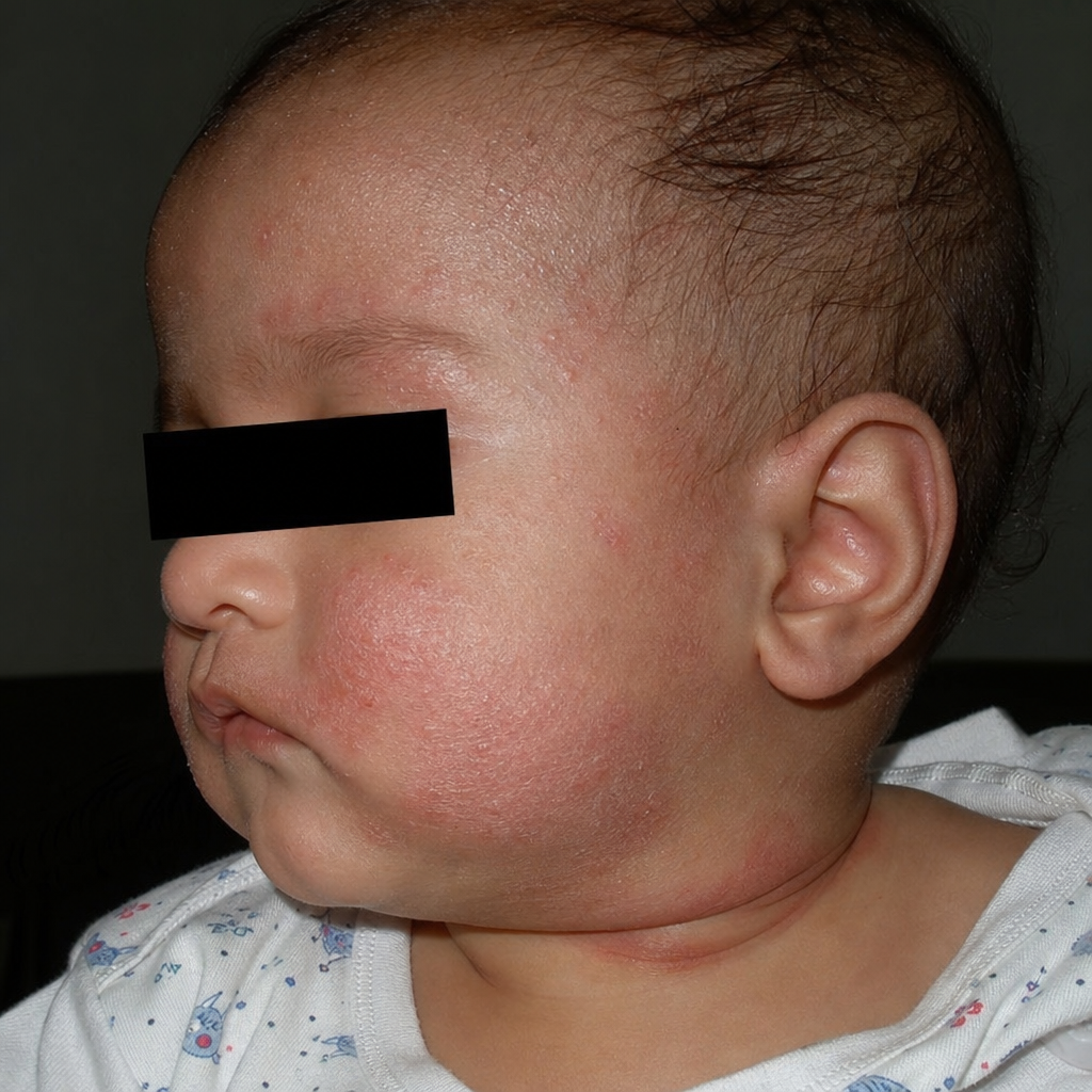
Koebner's phenomenon is seen in all except
The following is an important feature of psoriasis:
In which of the following conditions is the Koebner phenomenon most commonly observed?
Match the following scale types with their lesions. | Scales | Lesions | | :-- | :-- | | 1. Collarette scales | a. Pityriasis versicolour | | 2. Silvery scales | b. Pityriasis rosea | | 3. Mica-like scales | c. Psoriasis | | 4. Branny scales | d. Pityriasis lichenoides |
A 30-year-old male presented with silvery scales on elbow and knee, that bleed on removal. The probable diagnosis is:
All are nail changes seen in cases of psoriasis except:
A 54-year-old man presents with well-demarcated scaly plaques on the extensor surfaces of elbows and knees. The scales are silvery-white in appearance. What is the most likely diagnosis?
All are true about psoriasis except:
A patient with psoriasis who was started on systemic steroids develops generalized pustules all over the body after stopping treatment. What is the most likely cause?
Explanation: Secondary hyperparathyroidism - Calcitriol is the active form of vitamin D (1,25-dihydroxyvitamin D₃), and it is crucial for regulating calcium and phosphate levels in the body [1]. - In secondary hyperparathyroidism, often seen in chronic kidney disease (CKD), the kidneys cannot convert vitamin D to its active form, leading to hypocalcemia and increased PTH secretion [1], [2]. - Calcitriol supplementation helps to increase calcium absorption from the gut and suppress the release of parathyroid hormone (PTH), thereby treating the underlying cause of secondary hyperparathyroidism [1], [2]. - This is the primary therapeutic indication for calcitriol in clinical practice. Lichen planus - This is a chronic inflammatory condition affecting the skin, hair, nails, and mucous membranes - Typically treated with corticosteroids or other immunosuppressants - Calcitriol has no primary role in the treatment of lichen planus; its therapeutic applications are predominantly related to calcium and bone metabolism Pemphigus - Pemphigus is a group of rare autoimmune blistering diseases that affect the skin and mucous membranes - Primary treatment involves immunosuppressants like corticosteroids, often in high doses - Calcitriol is not indicated for the treatment of pemphigus, as its mechanism of action is unrelated to the autoimmune processes characteristic of this disease Leprosy - Leprosy is a chronic infectious disease caused by the bacterium Mycobacterium leprae - Treated with multi-drug therapy (MDT), which includes antibiotics like rifampicin, dapsone, and clofazimine - Calcitriol is not an antibiotic and therefore has no role in treating the bacterial infection responsible for leprosy
Explanation: ***Tinea corporis*** - **Koebner's phenomenon**, also known as the isomorphic response, is the appearance of skin lesions characteristic of a **pre-existing dermatosis** at sites of **trauma** to previously uninvolved skin. - **Tinea corporis**, a **superficial fungal infection**, does NOT exhibit true Koebner's phenomenon. - Its spread occurs through **direct fungal contact or autoinoculation**, not through an isomorphic response to non-specific trauma. *Psoriasis* - **Psoriasis** is the **classic example** of Koebner's phenomenon. - New psoriatic plaques can appear at sites of **skin trauma** such as scratches, surgical scars, burns, or tattoos within **10-20 days** of injury. - This occurs in approximately **25-50%** of psoriasis patients. *Warts* - **Warts** (verruca vulgaris), caused by **human papillomavirus (HPV)**, can show what is sometimes called **pseudo-Koebner's phenomenon**. - Trauma facilitates **viral inoculation** and seeding of HPV into the skin, leading to new wart formation along scratch lines. - However, this is technically **viral spread through trauma**, not a true isomorphic response of a pre-existing dermatosis. *Molluscum contagiosum* - **Molluscum contagiosum** can similarly demonstrate **pseudo-Koebner's phenomenon**. - Scratching spreads the **molluscum contagiosum virus** to adjacent areas, creating linear arrays of lesions. - Like warts, this represents **direct viral inoculation** rather than true isomorphic response, but is often grouped with Koebner's phenomenon in clinical practice.
Explanation: ***Silvery Scaling*** - **Silvery scaling** is a hallmark clinical feature of **psoriasis**, resulting from the rapid turnover of skin cells. - These scales often appear on **erythematous plaques** and can be easily scraped off, sometimes revealing pinpoint bleeding underneath (**Auspitz sign**). *Erythematous macules* - While psoriasis does involve **erythema** (redness), the primary lesions are typically **plaques**, not macules (flat, discolored spots). - Macules are seen in other dermatological conditions such as drug eruptions or early viral exanthems, but not as the definitive feature of psoriasis. *Crusting* - **Crusting** is a feature of conditions involving exudation and drying of serum, blood, or pus, such as **impetigo** or **eczema** with secondary infection. - It is not a characteristic primary lesion of psoriasis, although secondary infection of psoriatic plaques could theoretically lead to crusting. *Coarse bleeding* - **Coarse bleeding** is not a primary feature of psoriasis; however, when psoriatic scales are removed, pinpoint bleeding known as the **Auspitz sign** can occur. - This is distinct from frank, coarse bleeding and is a diagnostic clue rather than a characteristic lesion in itself.
Explanation: ***Correct: Psoriasis*** - **Psoriasis** is the **most classic and commonly cited example** of the Koebner phenomenon (isomorphic response) - New psoriatic plaques characteristically develop at sites of cutaneous trauma, scratches, or surgical incisions in 25-50% of psoriasis patients - This is a **pathognomonic feature** frequently tested in competitive exams and considered the prototype condition for demonstrating this phenomenon - The mechanism involves inflammatory cascades triggered by trauma in genetically predisposed skin *Incorrect: Lichen planus* - While lichen planus does exhibit the Koebner phenomenon with purplish polygonal papules appearing along scratch lines, it is **less commonly observed** compared to psoriasis - Seen in approximately 10-25% of lichen planus cases - Not considered the primary example when teaching about Koebner phenomenon *Incorrect: Viral warts* - Viral warts can demonstrate **pseudo-Koebner phenomenon** where new warts form along trauma lines due to viral inoculation - This is more accurately described as **autoinoculation** rather than true isomorphic response - Less commonly discussed in the context of classic Koebner phenomenon compared to psoriasis *Incorrect: All of the options* - While all three conditions can show Koebner-like responses, the question asks for "**most commonly observed**" - Psoriasis remains the **gold standard** and most frequently encountered example in clinical practice and medical literature
Explanation: ***1-b, 2-c, 3-d, 4-a*** - **Collarette scales** are pathognomonic of **Pityriasis rosea**, appearing as fine, trailing scales around the periphery of oval lesions in a "Christmas tree" distribution. - **Silvery scales** are the classic hallmark of **Psoriasis**, presenting as thick, adherent, silvery-white scales overlying well-demarcated erythematous plaques. - **Mica-like scales** are characteristic of **Pityriasis lichenoides**, appearing as thick, shiny, adherent scales that can be peeled off like mica sheets. - **Branny scales** are typical of **Pityriasis versicolor**, presenting as fine, powdery scales caused by **Malassezia** yeast overgrowth. *1-d, 2-c, 3-a, 4-b* - Incorrectly matches **collarette scales with Pityriasis lichenoides**, which typically presents with mica-like scales, not collarette scales. - Misassociates **mica-like scales with Pityriasis versicolor**, which characteristically has branny (fine, powdery) scales. *1-c, 2-b, 3-d, 4-a* - Wrongly pairs **collarette scales with Psoriasis**, which is known for thick silvery scales, not peripheral collarette scales. - Incorrectly matches **silvery scales with Pityriasis rosea**, which has collarette scales at lesion periphery, not silvery scales. *1-a, 2-b, 3-d, 4-c* - Falsely associates **collarette scales with Pityriasis versicolor**, which has branny scales from yeast infection, not collarette scales. - Mismatches **branny scales with Psoriasis**, which has characteristic thick silvery scales, not fine powdery scales.
Explanation: ***Psoriasis*** - The presence of **silvery scales** on the elbows and knees, which **bleed upon removal** (Auspitz sign), is a classic presentation of **plaque psoriasis**. - Psoriasis is a chronic inflammatory skin condition characterized by **accelerated epidermal turnover**. *Secondary syphilis* - Secondary syphilis typically presents with a **generalized maculopapular rash**, which can affect the palms and soles, but it does not usually feature silvery scales or the Auspitz sign. - Other common symptoms of secondary syphilis include **fever, lymphadenopathy, and condyloma lata**. *Pityriasis* - **Pityriasis rosea** is characterized by an oval, fawn-colored, scaly rash, often preceded by a **herald patch**, and usually resolves spontaneously. It does not typically present with silvery scales or bleeding on removal. - **Pityriasis versicolor** is caused by yeast and presents as hypopigmented or hyperpigmented macules with fine scales, commonly on the trunk, not silvery scales on elbows and knees. *Seborrhoeic dermatitis* - Seborrhoeic dermatitis involves greasy, yellowish scales on red skin, typically affecting areas rich in sebaceous glands like the scalp, face (nasolabial folds, eyebrows), and chest. - It does not present with silvery scales or the Auspitz sign, which are specific to psoriasis.
Explanation: ***Mees lines*** - **Mees lines** (or Aldrich-Mees lines) are **transverse white bands** that appear in the nail plate. - They are typically associated with **heavy metal poisoning** (e.g., arsenic), chemotherapy, or systemic illnesses, not psoriasis. *Subungual hyperkeratosis* - This is a common finding in **psoriasis**, characterized by the **thickening of the nail bed** due to excessive keratin production. - It leads to lifting of the nail plate from the nail bed. *Oil drop sign* - The **oil drop sign** (or salmon patch) is a classic psoriatic nail change, presenting as a **translucent, yellowish-red discoloration** under the nail plate. - It is due to psoriasis of the nail bed. *Pitting* - **Nail pitting** refers to the presence of **small depressions or pits** on the nail surface. - It results from defective keratinization of the nail matrix and is a characteristic sign of nail psoriasis.
Explanation: ***Psoriasis*** - **Psoriasis** classically presents with **scaly plaques** that often appear on the **extensor surfaces** like elbows and knees. - The scales are typically **silvery-white** and can be associated with itching or discomfort. *Eczema* - **Eczema** (atopic dermatitis) typically presents with **pruritic (itchy)**, **erythematous (red)**, and sometimes **weeping lesions**, often in flexural areas like the antecubital and popliteal fossae. - While it can be scaly, the scales are usually finer and less prominent than those seen in psoriasis, and it often presents on **flexor surfaces**. *Lichen planus* - **Lichen planus** is characterized by **pruritic, purple, polygonal, planar papules and plaques** (the "6 Ps"), often found on flexural surfaces, wrists, and ankles. - It does not typically present with the thick, silvery scales on extensor surfaces seen in this case. *Pityriasis rosea* - **Pityriasis rosea** typically begins with a **herald patch**, followed by an eruption of smaller, oval, scaly patches that follow the skin Langer's lines, often described as a "Christmas tree" pattern on the trunk. - It is usually self-limiting and rarely involves the thick, scaly plaques on extensor surfaces seen in psoriasis.
Explanation: ***Very pruritic*** - While psoriasis can be itchy, it is generally not characterized as "very pruritic" compared to other dermatological conditions like **eczema** or **scabies**. - **Pruritus** in psoriasis tends to be mild to moderate, and it is not a defining characteristic that differentiates it from other skin disorders. *Parakeratosis & acanthosis* - **Parakeratosis** (retention of nuclei in the stratum corneum) and **acanthosis** (epidermal hyperplasia) are classic histopathological hallmarks of psoriasis. - These features reflect the rapid epidermal turnover characteristic of the condition. *Pitting of nails* - **Nail pitting**, onycholysis, and subungual hyperkeratosis are common and characteristic manifestations of psoriasis, affecting up to 50% of patients. - These nail changes are highly indicative of **psoriatic involvement**. *Joint involvement in 5–10%* - **Psoriatic arthritis**, involving inflammation of the joints, affects approximately 5-30% of individuals with psoriasis. - This statistic makes joint involvement a significant comorbidity of psoriasis.
Explanation: ***Pustular psoriasis due to steroid withdrawal*** - **Systemic steroid withdrawal** can precipitate a severe flare of psoriasis, often leading to generalized **pustular psoriasis**. - This is a well-known phenomenon where the suppression of the immune system by steroids is abruptly removed, causing a rebound inflammatory response. *Drug-induced pustular psoriasis* - While certain drugs can induce pustular psoriasis, the scenario specifically highlights the **cessation of systemic steroids** as the trigger. - This option doesn't pinpoint the direct causal effect of stopping the medication. *Bacterial infection* - Although pustules can be associated with bacterial infections, the **generalized nature** and history of **steroid withdrawal** in a patient with psoriasis make an infectious cause less likely as the primary trigger. - A bacterial infection would typically present with signs of local infection (e.g., warmth, tenderness, fever) alongside the pustules, which are not exclusively mentioned here. *Septicemia* - **Septicemia** is a severe bloodstream infection and would present with systemic signs of illness such as high fever, chills, hypotension, and organ dysfunction, which are not described. - While pustules can sometimes occur in severe infections, the clinical context strongly points to a dermatological reaction to medication changes, not a systemic infection.
Get full access to all questions, explanations, and performance tracking.
Start For Free