
Psoriasis — MCQs

On this page
Koebner phenomenon is seen in which one of the following conditions?
A 45-year-old man presents with a 6-month history of scaly, erythematous plaques with silvery scales on his elbows, knees, and scalp. He reports occasional joint pain. His sister has similar skin problems. Examination reveals well-demarcated, erythematous plaques covered with silvery scales. Removal of scales causes pinpoint bleeding. Which of the following is most likely to be elevated in this condition?
Typical silvery scales of psoriasis are absent in –
A patient with psoriasis was started on systemic steroids. After stopping the treatment, the patient developed universally red scaly skin with plaques losing their margins all over his body. The most likely cause is –
Silver plaques are a feature of:
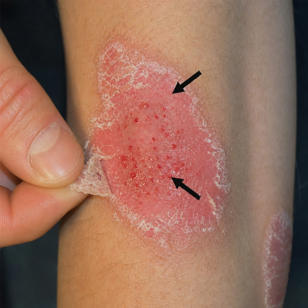
On removal of scales from a psoriatic lesion, the sign shown (indicated by arrows) is:
All are true about psoriasis except –
What is the most common type of psoriasis?
Dithranol ointment is used for:
All are nail changes seen in cases of psoriasis except:
Practice by Chapter
Pathophysiology of Psoriasis
Practice Questions
Psoriasis Vulgaris
Practice Questions
Guttate Psoriasis
Practice Questions
Erythrodermic Psoriasis
Practice Questions
Pustular Psoriasis
Practice Questions
Palmoplantar Psoriasis
Practice Questions
Nail Psoriasis
Practice Questions
Scalp Psoriasis
Practice Questions
Psoriatic Arthritis
Practice Questions
Topical Therapy for Psoriasis
Practice Questions
Systemic Therapy for Psoriasis
Practice Questions
Phototherapy and Biologics for Psoriasis
Practice Questions
Want unlimited practice?
Get full access to all questions, explanations, and performance tracking.
Scan to download app