
Pediatric Dermatology — MCQs

On this page
Which of the following conditions produces seborrheic dermatitis-like lesions in an infant?
Which of the following is a characteristic skin lesion of Tuberous sclerosis?
What is the earliest presenting feature of tuberous sclerosis?
Which of the following skin lesions does not change or remains the same throughout life?
Which of the following statements is NOT true about Sturge-Weber syndrome?
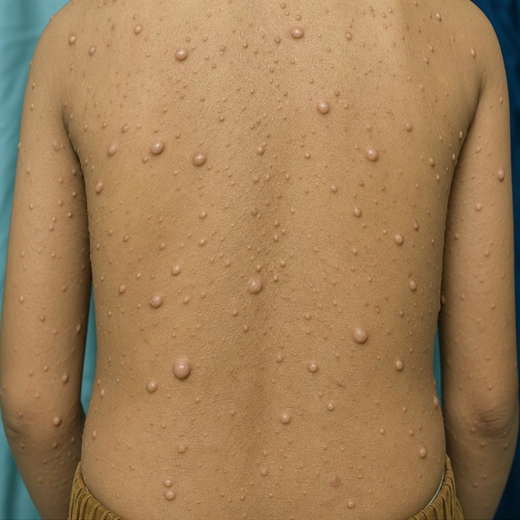
These lesions are seen in which condition?
All of the following are true about dyskeratosis congenita, EXCEPT:
A 10-year-old boy presents with multiple tan-colored patches on his skin and freckle-like changes in his axillary area. The remainder of his clinical examination is normal. Which of the following conditions is also found in patients with this disorder as they age?
Which of the following is not a vascular malformation?
Which of the following is NOT included under the diagnostic criteria for Neurofibromatosis type 1?
Practice by Chapter
Neonatal Dermatology
Practice Questions
Infantile Hemangiomas and Vascular Malformations
Practice Questions
Atopic Dermatitis in Children
Practice Questions
Acne in Childhood and Adolescence
Practice Questions
Childhood Exanthems
Practice Questions
Genetic Skin Disorders in Children
Practice Questions
Genodermatoses
Practice Questions
Nutritional Dermatoses in Children
Practice Questions
Pigmentary Disorders in Children
Practice Questions
Hair Disorders in Children
Practice Questions
Child Abuse: Cutaneous Manifestations
Practice Questions
Therapeutic Considerations in Pediatric Dermatology
Practice Questions
Want unlimited practice?
Get full access to all questions, explanations, and performance tracking.
Scan to download app