Which of the following is not considered an opportunistic infection in AIDS?
A farmer presents you with a cauliflower-shaped mass on foot, which developed after a minor injury. Microscopy shows copper penny bodies. What is the most likely diagnosis?
A 45-year-old patient with a history of poorly controlled diabetes presents with sinus pain, nasal discharge, and facial swelling. A biopsy reveals broad, nonseptate hyphae branching at wide angles. What is the most likely causative agent?
Fungal infection which is acquired by traumatic inoculation is?
A 65-year-old diabetic man presents with black necrotic tissue on his palate. What is the most likely causative organism?
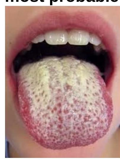
An HIV positive patient with a CD4 count of 300/cumm presents with mucosal lesions in the mouth as shown in the figure. On microscopy, budding yeasts and pseudohyphae are seen. A most probable diagnosis is?

A patient presents with annular, scaly plaques with perifollicular extension on the trunk. What is the most likely diagnosis?
A 24 year old man had multiple, small hypopigmented macules on the upper chest and back for the last three months. The macules were circular, arranged around follicles and many had coalesced to form large sheets. The surface of the macules showed fine scaling. He had similar lesions one year ago which subsided with treatment. The most appropriate investigation to confirm the diagnosis is -
A child comes with a circular 3cm x 3cm scaly patchy hair loss with itching in the lesions. The investigation of choice is
Which of the following tests is used in the diagnosis of tinea faciei?
Explanation: ***Rubella*** - Rubella, or **German measles**, is a relatively mild viral infection that typically affects children and is not considered an **opportunistic infection** in immunocompromised individuals like those with AIDS [1]. - While it can cause congenital rubella syndrome in infants whose mothers are infected during pregnancy, it does not disproportionately affect or cause severe disease in AIDS patients due to their compromised immunity [1]. *Candidiasis* - **Oropharyngeal** and **esophageal candidiasis** are common opportunistic infections in AIDS patients, often indicating significant immune suppression [2,3]. - The fungus *Candida albicans* can proliferate unchecked when the **CD4 count** is low [2]. *Kaposi's sarcoma* - This is a **cancer** caused by the **human herpesvirus 8 (HHV-8)**, which is a classic AIDS-defining illness [3]. - Its presence indicates severe immunodeficiency and was a hallmark of the early AIDS epidemic [3]. *Cytomegalovirus infection* - **Cytomegalovirus (CMV)** can cause severe and widespread disease in AIDS patients, including **retinitis**, **colitis**, and **encephalitis** [2]. - It becomes a significant risk when the **CD4 count** drops below 100 cells/mm³ [2].
Explanation: **Chromoblastomycosis** - The characteristic "cauliflower-shaped" lesion on the foot following a minor injury, especially in a farmer (indicating outdoor exposure), is highly suggestive of chromoblastomycosis. - The presence of **copper penny bodies** (also known as **sclerotic** or **muriform cells**) on microscopy is **pathognomonic** for chromoblastomycosis. *Blastomycosis* - Blastomycosis typically presents with **granulomatous lesions** that can ulcerate but are rarely described as cauliflower-shaped. - Microscopic examination would reveal **broad-based budding yeast cells**, not copper penny bodies. *Sporotrichosis* - Sporotrichosis usually presents as **subcutaneous nodules** that can ulcerate and spread lymphatically, forming a chain of lesions. - Microscopy shows **cigar-shaped budding yeasts** within macrophages or neutrophils, which are distinct from copper penny bodies. *Phaeohyphomycosis* - Phaeohyphomycosis encompasses a broad group of infections by dematiaceous fungi that produce **dark-walled hyphae** or yeast-like cells in tissue. - While it can cause subcutaneous nodules or cysts, the presence of distinct copper penny bodies points away from phaeohyphomycosis as the primary diagnosis.
Explanation: ***Rhizopus spp.*** - The patient's presentation with **sinus pain**, **nasal discharge**, **facial swelling**, and a history of **poorly controlled diabetes** is classic for **mucormycosis** (also known as zygomycosis). - **Biopsy findings** of **broad, nonseptate hyphae branching at wide/irregular angles** are pathognomonic for mucormycosis, most commonly caused by *Rhizopus* species. - Diabetes mellitus, particularly when poorly controlled with **ketoacidosis**, is a major risk factor for rhinocerebral mucormycosis. *Aspergillus fumigatus* - This fungus typically causes infections with **septate hyphae** that **branch at acute angles** (45 degrees), which is morphologically distinct from mucormycosis. - While *Aspergillus* can cause invasive sinusitis in immunocompromised patients, the specific hyphal morphology (nonseptate, wide-angle branching) points away from it. *Candida albicans* - *Candida albicans* is a yeast that typically appears as **oval budding cells** and **pseudohyphae** on microscopy, not broad, nonseptate hyphae. - While it can cause opportunistic infections in diabetic and immunocompromised patients, its microscopic appearance is entirely inconsistent with the biopsy findings. *Cryptococcus neoformans* - *Cryptococcus neoformans* is an **encapsulated yeast** that is typically identified by its **spherical or oval budding cells** with a characteristic **polysaccharide capsule** visible with India ink stain. - It primarily causes **meningitis** and pulmonary infections in immunocompromised hosts, and its morphology (yeast, not hyphae) is entirely different from the described findings.
Explanation: ***Sporothrix*** - **Sporotrichosis** is characteristically acquired through **traumatic inoculation** of the fungus, often from contact with soil, thorns, or decaying vegetation. - The organism causes **cutaneous lymphatic disease**, presenting as nodular lesions along lymphatic drainage paths. *Blastomyces* - **Blastomycosis** is typically acquired by inhaling airborne fungal spores, usually from **soil rich in organic matter** or decaying wood. - It primarily affects the **lungs** and can disseminate to the skin, bones, and other organs, but is not primarily associated with traumatic inoculation. *Coccidioides* - **Coccidioidomycosis** (Valley Fever) is acquired by inhaling **arthroconidia** present in dust or soil in endemic areas. - It is a **pulmonary infection** that can disseminate to other body sites, and its entry is almost exclusively respiratory, not traumatic. *Paracoccidioides* - **Paracoccidioidomycosis** is acquired by inhaling airborne fungal propagules, typically found in **soil in Latin America**. - It primarily causes **chronic pulmonary disease** and can spread to mucous membranes, skin, and lymph nodes, with no known association with traumatic inoculation.
Explanation: ***Mucor species*** - The presence of **black necrotic tissue** on the palate in a diabetic patient is highly suggestive of **mucormycosis**, an aggressive fungal infection caused by *Mucor* species. - **Diabetes mellitus**, particularly with ketoacidosis, is a major risk factor for mucormycosis due to impaired phagocytic function and increased iron availability. *Cryptococcus neoformans* - This fungus is primarily associated with **cryptococcal meningitis** or pneumonia, especially in immunocompromised individuals. - It does not typically cause **black necrotic lesions** on the palate. *Candida albicans* - While *Candida albicans* can cause oral infections (**thrush**), these typically present as white, creamy patches that can be scraped off, not black necrotic tissue. - Oral candidiasis is common in diabetics but does not usually involve tissue necrosis. *Aspergillus fumigatus* - *Aspergillus* species can cause invasive infections, particularly in immunocompromised patients, often affecting the lungs or sinuses. - While it can cause **necrotic lesions**, the characteristic rapid progression and specific presentation in the palate of a diabetic with black necrotic tissue points more strongly towards *Mucor*.
Explanation: ***Candidiasis*** - The image shows **white, creamy patches** on the tongue, which are characteristic of **oral candidiasis** (thrush). These lesions are often easily **scrapable**. - The presence of **budding yeasts and pseudohyphae** on microscopy confirms a fungal infection, and in an **HIV-positive patient with a CD4 count of 300/cumm**, Candida infection is very common due to immunosuppression. *Hairy leukoplakia* - Hairy leukoplakia presents as **white, corrugated, non-scrapable lesions**, typically on the lateral borders of the tongue. - It is caused by the **Epstein-Barr virus (EBV)** and does not show budding yeasts or pseudohyphae on microscopy. *Lichen planus* - Oral lichen planus presents with **white, lacy patterns (Wickham's striae)** on the buccal mucosa or tongue, which are usually not scrapable. - It is a **chronic inflammatory condition** and not an infectious process characterized by yeasts and pseudohyphae. *Diphtheria* - Diphtheria causes the formation of a **thick, gray pseudomembrane** in the throat and tonsils, which is firmly adherent and can cause bleeding if removed. - It is a **bacterial infection** caused by *Corynebacterium diphtheriae*, and microscopic examination would reveal characteristic gram-positive rods, not yeasts.
Explanation: ***Tinea*** - **Tinea corporis** classically presents with **annular, scaly plaques with central clearing** and an active, raised border. - On hairy areas or with follicular involvement, dermatophyte infections show **perifollicular extension** as the fungus invades hair follicles. - The **annular morphology with scale** is pathognomonic for dermatophyte infection, confirmed by **KOH preparation** showing septate hyphae. - Common sites include trunk, limbs, and any body area with hair follicles. *Psoriasis* - Presents with **well-demarcated, erythematous plaques** with **silvery-white scales**, typically on extensor surfaces (elbows, knees, scalp). - **Follicular psoriasis** is rare and shows **pinpoint follicular papules**, not annular plaques with perifollicular extension. - Auspitz sign (pinpoint bleeding on scale removal) helps differentiate from tinea. *Lichen planus* - Characterized by **pruritic, polygonal, purple, planar papules** (the "6 Ps"). - **Lichen planopilaris** (follicular variant) causes **scarring alopecia** with follicular hyperkeratosis, not annular scaly plaques. - Wickham striae may be visible on mucosal surfaces. *Pityriasis versicolor* - Caused by **Malassezia species**, presents as **hypo- or hyperpigmented macules** with fine scale on trunk and upper arms. - **Follicular variant** (pityriasis folliculorum) shows discrete follicular papules, NOT annular plaques. - "Spaghetti and meatballs" appearance on KOH prep (short hyphae and spores) differentiates from dermatophytes.
Explanation: ***Potassium hydroxide preparation of scales*** - The description of **hypopigmented macules** with **fine scaling** on the upper chest and back, which coalesced and recurred, is highly suggestive of **Pityriasis versicolor** (also known as Tinea versicolor). - A **potassium hydroxide (KOH) preparation** of the scales directly visualizes the fungal elements (**hyphae and spores**, often described as "spaghetti and meatballs" appearance), confirming the diagnosis. *Slit skin smear from discrete macules* - A **slit skin smear** is primarily used for diagnosing mycobacterial infections, particularly **leprosy**, to detect acid-fast bacilli. - This procedure is not appropriate for diagnosing superficial fungal infections like Pityriasis versicolor. *Skin biopsy of coalesced macules* - While a **skin biopsy** can show fungal elements in the stratum corneum, it is an invasive and generally unnecessary procedure for diagnosing Pityriasis versicolor. - **KOH preparation** is a faster, simpler, and less invasive method that provides a definitive diagnosis. *Tzanck test* - The **Tzanck test** is used to identify multinucleated giant cells characteristic of viral infections, such as **herpes simplex virus** or **varicella-zoster virus**. - It involves scraping the base of a vesicle or bulla, which is not consistent with the described presentation of scaling macules.
Explanation: ***Correct: KOH mount (Potassium Hydroxide mount)*** - A **KOH mount** is the investigation of choice for suspected **dermatophyte infections** (tinea capitis), which commonly present as circular, scaly patches of hair loss with itching in children. - It involves dissolving keratinous material to visualize **fungal hyphae** and spores directly under a microscope. - This is a quick, cost-effective, and highly specific first-line diagnostic test. *Incorrect: Tzanck smear* - A **Tzanck smear** is primarily used to diagnose **viral infections** like herpes simplex or varicella-zoster by identifying multinucleated giant cells. - It is not effective for detecting fungal elements responsible for scaly hair loss. *Incorrect: Gram stain* - A **Gram stain** is a technique used to classify **bacteria** based on their cell wall properties. - It would not reveal fungal hyphae or spores relevant to the described condition. *Incorrect: Split skin smear* - A **split skin smear** (or slit-skin smear) is typically used in the diagnosis of **leprosy** to identify acid-fast bacilli. - This technique involves scraping the dermis and is not suitable for diagnosing superficial fungal infections.
Explanation: ***KOH mount*** - A **KOH (potassium hydroxide) mount** is the gold standard for diagnosing **dermatophyte infections**, including **tinea faciei**. - The KOH solution dissolves keratinocytes, allowing for the visualization of fungal **hyphae and spores** under a microscope. *Gram's stain* - **Gram's stain** is primarily used to differentiate bacterial species based on their cell wall properties. - It does not effectively visualize **fungal elements** and is therefore not used for diagnosing tinea infections. *Tissue smear* - A **tissue smear** involves examining cells from a lesion, typically for conditions like viral infections (e.g., Tzanck smear for herpes) or some cutaneous malignancies. - It is not the standard or preferred method for identifying **dermatophyte fungal structures**. *Wood's lamp* - A **Wood's lamp**, which emits ultraviolet light, is used to detect certain fungal infections (e.g., *Microsporum* species causing tinea capitis) and bacterial conditions (e.g., erythrasma). - Many common dermatophytes causing **tinea faciei** (e.g., *Trichophyton*) do not fluoresce under a Wood's lamp, making it an unreliable diagnostic tool for this specific condition.
Get full access to all questions, explanations, and performance tracking.
Start For Free