
Dermatitis and Eczema — MCQs

On this page
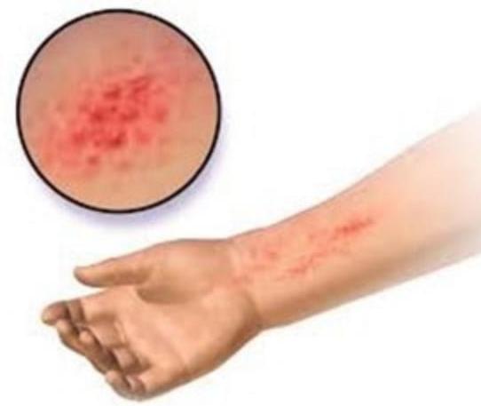
A child has a rash. His family history is positive for asthma. What could be the most probable diagnosis?
Most common flexural site for atopic dermatitis -
Which of the following organisms has a role to play in Seborrheic dermatitis?
Characteristic of chronic eczema?
Most common metal in contact allergic dermatitis is?
A 25-year-old patient presents with chronic itchy, erythematous skin lesions on the flexural areas that have been recurring since childhood. The patient has a family history of asthma. Which of the following is the most important diagnostic criterion for the most likely diagnosis?
Which of the following is NOT a feature of atopic dermatitis?
A primigravida female presented with erythematous skin changes and lesions consistent with a severe allergic reaction. Which of the following should be the most appropriate treatment?
Which condition is characterized by perioral pallor and Dennie-Morgan folds?
Most common precipitant of contact dermatitis is?
Practice by Chapter
Atopic Dermatitis
Practice Questions
Seborrheic Dermatitis
Practice Questions
Contact Dermatitis: Irritant
Practice Questions
Contact Dermatitis: Allergic
Practice Questions
Nummular Eczema
Practice Questions
Dyshidrotic Eczema
Practice Questions
Stasis Dermatitis
Practice Questions
Asteatotic Eczema
Practice Questions
Lichen Simplex Chronicus
Practice Questions
Autoeczematization (Id Reaction)
Practice Questions
Photosensitive Eczemas
Practice Questions
Treatment Strategies for Eczematous Disorders
Practice Questions
Want unlimited practice?
Get full access to all questions, explanations, and performance tracking.
Scan to download app