
Dermatitis and Eczema — MCQs

On this page
A 5-year-old boy presents with a red, itchy rash on his cheeks and the flexural surfaces of his limbs. What is the likely diagnosis?
A patient presents with erythematous, well-demarcated plaques with greasy scales on the scalp and central face. What is the most likely diagnosis?
A 35-year-old woman presents with erythematous, scaly plaques in the nasolabial folds, eyebrows, and scalp. What is the most likely diagnosis?
A 5-year-old child presents with intense itching and erythematous papules on the extensor surfaces of the arms and legs. What is the most likely diagnosis?
A 40-year-old female presents with a pruritic rash on her hands after using a new soap. Patch testing shows a positive reaction to the fragrance mix. What is the most appropriate management?
A 27-year-old woman presents with a painful blistering rash on her right shoulder after a hiking trip. She reports recent exposure to poison ivy. What is the most likely diagnosis?
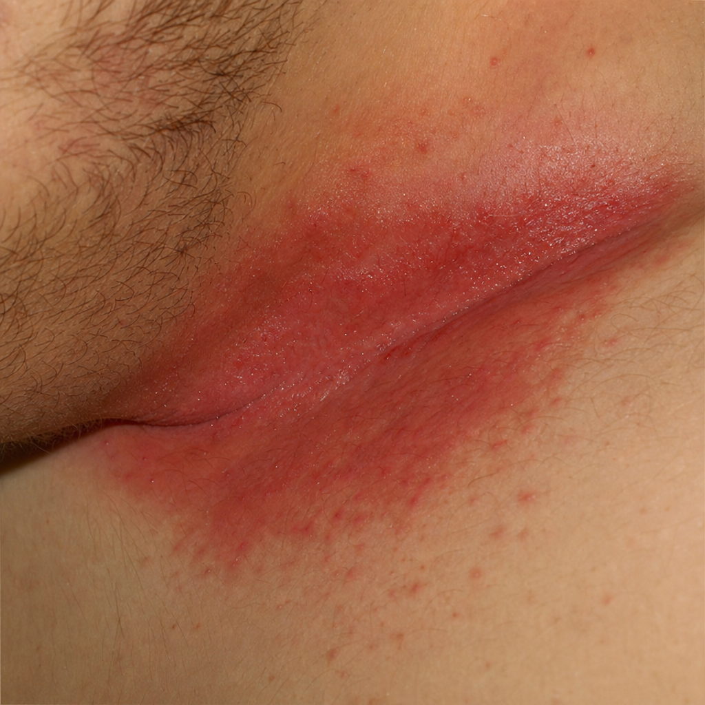
Identify the skin condition shown in the image.
What condition is likely to be present in the child shown in the image, whose mother has asthma?
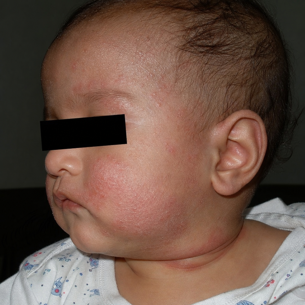
What condition is likely to develop in a child with a mother who has asthma?
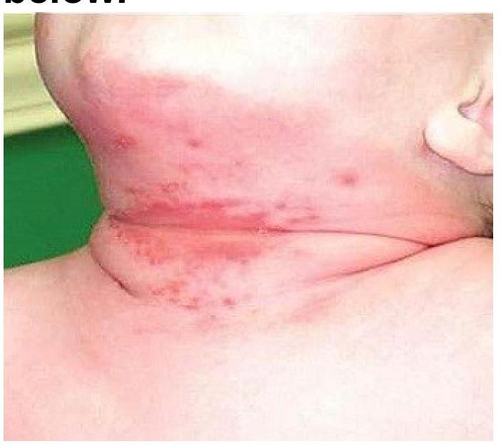
A child presents with a rash in the neck folds as shown in the image. The area appears erythematous with satellite lesions and maceration. What is the most likely diagnosis?
Practice by Chapter
Atopic Dermatitis
Practice Questions
Seborrheic Dermatitis
Practice Questions
Contact Dermatitis: Irritant
Practice Questions
Contact Dermatitis: Allergic
Practice Questions
Nummular Eczema
Practice Questions
Dyshidrotic Eczema
Practice Questions
Stasis Dermatitis
Practice Questions
Asteatotic Eczema
Practice Questions
Lichen Simplex Chronicus
Practice Questions
Autoeczematization (Id Reaction)
Practice Questions
Photosensitive Eczemas
Practice Questions
Treatment Strategies for Eczematous Disorders
Practice Questions
Want unlimited practice?
Get full access to all questions, explanations, and performance tracking.
Scan to download app