
Dermatitis and Eczema — MCQs

On this page
A child has a pruritic rash as shown below. His mother is an asthmatic. Comment on the diagnosis:
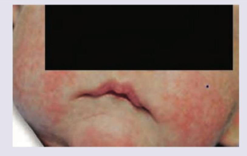
A one-year-old child presents with the following lesion on the face. His mother has a history of bronchial asthma. What is the diagnosis?
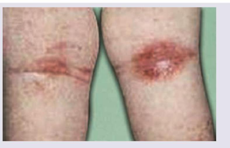
A child presented with itchy plaques over the neck, the bilateral popliteal and cubital fossa. What could be the diagnosis?
Dennie-Morgan fold is indicative of:
A patient comes to you with skin reactions after visiting the hair dresser. What will you do to confirm the diagnosis of contact dermatitis?
A 70-year-old man comes to the emergency department because of a skin rash and severe itching. He appears ill; there is a generalized skin rash that is scaly, erythematous, and thickened. His palms, soles, and scalp are also involved. Which of the following is the most likely diagnosis?
A patient with atopic dermatitis shows the following findings. Which indicates POOR prognosis? 1. Onset before age 2 2. Flexural involvement 3. Palmar hyperlinearity 4. Filaggrin mutation
A patient with atopic dermatitis shows poor response to topical steroids. Next best step is:
A 45-year-old woman with poorly controlled type 2 diabetes (HbA1c 9.2%) and chronic kidney disease presents with intensely pruritic nodules on extensor surfaces for 8 months. Previous treatments with topical steroids, antihistamines, and gabapentin provided minimal relief. Skin biopsy shows acanthosis, compact orthokeratosis, and increased nerve fibers. CBC shows eosinophilia. Most appropriate next step is:
Topical steroids are most effective in:
Practice by Chapter
Atopic Dermatitis
Practice Questions
Seborrheic Dermatitis
Practice Questions
Contact Dermatitis: Irritant
Practice Questions
Contact Dermatitis: Allergic
Practice Questions
Nummular Eczema
Practice Questions
Dyshidrotic Eczema
Practice Questions
Stasis Dermatitis
Practice Questions
Asteatotic Eczema
Practice Questions
Lichen Simplex Chronicus
Practice Questions
Autoeczematization (Id Reaction)
Practice Questions
Photosensitive Eczemas
Practice Questions
Treatment Strategies for Eczematous Disorders
Practice Questions
Want unlimited practice?
Get full access to all questions, explanations, and performance tracking.
Scan to download app