
Dermatitis and Eczema — MCQs

On this page
The Hanifin & Rajka diagnostic criteria are used for which condition?
A 17-year-old female presents with a pruritic rash localized to the wrist. Papules and vesicles are noted in a bandlike pattern, with slight oozing from some lesions. What is the most likely cause of this rash?
A 25-year-old male presents with recurrent episodes of flexural eczema, contact urticaria, recurrent skin infections, and severe abdominal cramps and diarrhea upon consuming seafood. What is the most likely diagnosis?
Air-borne contact dermatitis can be diagnosed by?
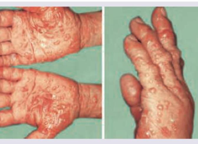
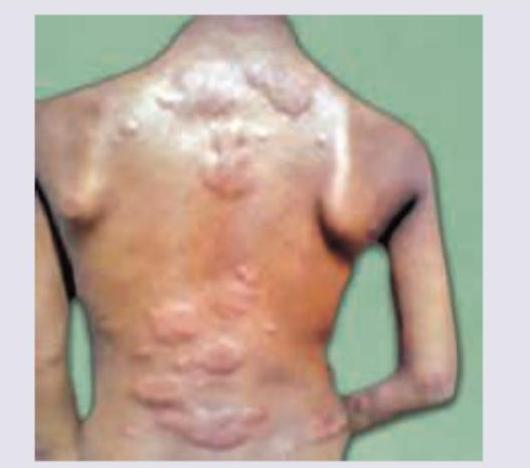
The following are true regarding the picture shown except:
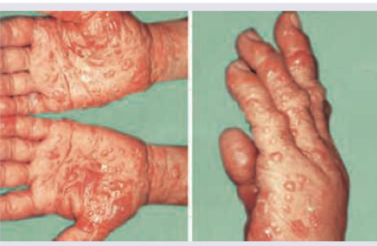
All are true about the lesions shown except:
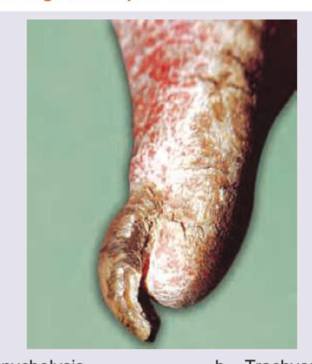
The image shows:
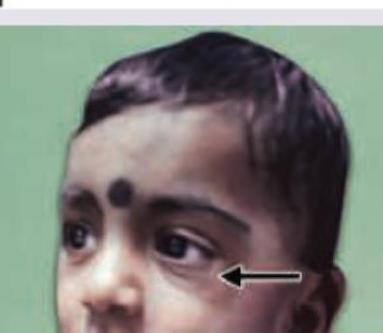
The clinical sign shown in the image is:
This patient was cleaning the basement, following which he developed the lesions as shown on his back for the last 6 hours. The image shows presence of:
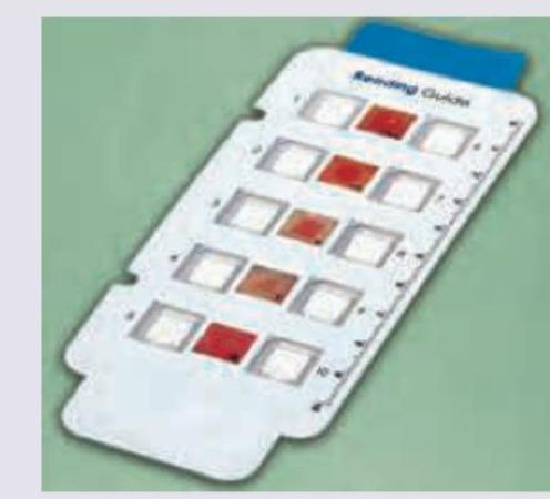
The following strip is used for testing of:
Practice by Chapter
Atopic Dermatitis
Practice Questions
Seborrheic Dermatitis
Practice Questions
Contact Dermatitis: Irritant
Practice Questions
Contact Dermatitis: Allergic
Practice Questions
Nummular Eczema
Practice Questions
Dyshidrotic Eczema
Practice Questions
Stasis Dermatitis
Practice Questions
Asteatotic Eczema
Practice Questions
Lichen Simplex Chronicus
Practice Questions
Autoeczematization (Id Reaction)
Practice Questions
Photosensitive Eczemas
Practice Questions
Treatment Strategies for Eczematous Disorders
Practice Questions
Want unlimited practice?
Get full access to all questions, explanations, and performance tracking.
Scan to download app