
Dermatitis and Eczema — MCQs

On this page
Which of the following is a minor clinical feature in the diagnosis of atopic dermatitis?
A child presents with itchy skin lesions on the elbows and other extensor surfaces. The mother reports a history of bronchial asthma. What is the most likely diagnosis?
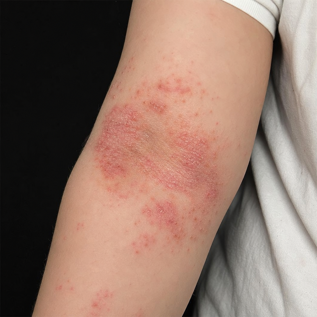
The day after playing in the high grasses of a neighbour's fields, an 8-year-old boy develops a weeping, vesicular, erythematous, and itchy rash on his arms, legs, and neck. Which of the following best describes this inflammatory response?
A 36-year-old farmer presented to the dermatology department with pruritic erythematous lesions on the arms, forearms, face, and retroauricular area after removing weeds in his farm. A diagnosis of phytodermatitis was made. What is the most likely plant responsible for this condition?
Which of the following materials used to make jewelry will commonly cause contact dermatitis in sensitive individuals?
A patient presents with a rash. There is a family history of asthma. What is the most probable diagnosis?
A 55-year-old male, with uncontrolled diabetes mellitus and hypertension, developed severe airborne contact dermatitis. What is the most appropriate drug for his treatment?
Id reaction is associated with which of the following conditions?
A 25-year-old male presents with multiple erythematous annular plaques with peripheral collarette of scales arranged predominantly over the trunk. What is the most probable diagnosis?
A 5-year-old boy presents with a small hypopigmented scaly macule on his cheek. Some of his classmates also have similar lesions. What is the most probable diagnosis?
Practice by Chapter
Atopic Dermatitis
Practice Questions
Seborrheic Dermatitis
Practice Questions
Contact Dermatitis: Irritant
Practice Questions
Contact Dermatitis: Allergic
Practice Questions
Nummular Eczema
Practice Questions
Dyshidrotic Eczema
Practice Questions
Stasis Dermatitis
Practice Questions
Asteatotic Eczema
Practice Questions
Lichen Simplex Chronicus
Practice Questions
Autoeczematization (Id Reaction)
Practice Questions
Photosensitive Eczemas
Practice Questions
Treatment Strategies for Eczematous Disorders
Practice Questions
Want unlimited practice?
Get full access to all questions, explanations, and performance tracking.
Scan to download app