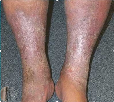
What is the most likely cause of these skin changes?

Pompholyx affects which area(s)?
All of the following are exogenous eczema except?
All are skin findings in atopic dermatitis, EXCEPT:
A 15-year-old healthy boy presents with pruritic, erythematous, blocky skin lesions on his arms, legs, and trunk that appear within an hour of consuming seafood. What is the most likely diagnosis?
Pityriasis rosea is best described as:
A 30-year-old male presents with pruritic, flat-topped, polygonal, shiny violaceous papules with flexural distribution. What is the most likely diagnosis?
A 27-year-old male presents with a 3-year history of itchy, excoriated papules on the forehead and exposed parts of the arms and legs. The condition is most severe during the rainy season and completely improves in winter. What is the most likely diagnosis?
What is the best initial treatment for acute contact dermatitis?
Which of the following causes allergic contact dermatitis through air?
Explanation: ***Chronic venous stasis*** - The image shows **hyperpigmentation**, **edema**, and **varicose veins** on the lower leg, which are classic features of **stasis dermatitis**. - Chronic venous insufficiency leads to **hemosiderin deposition** causing brown discoloration and **chronic inflammation** in the lower extremities. *Asteatotic eczema* - Typically presents with **dry, scaly, cracked skin** resembling a "crazy paving" pattern, not the hyperpigmented changes seen here. - Usually affects elderly patients and involves **xerosis** (dry skin) rather than **venous congestion** signs. *Necrobiosis lipoidica* - Characterized by **yellow-brown plaques** with **waxy appearance** and **telangiectasias**, typically on the shins. - Strongly associated with **diabetes mellitus** and shows **central atrophy** with raised borders, unlike the diffuse changes shown. *Pretibial myxedema* - Presents as **non-pitting, waxy, orange-peel textured plaques** on the anterior shins in **hyperthyroid patients**. - Associated with **Graves' disease** and lacks the **venous congestion** and **hemosiderin staining** visible in this case.
Explanation: **Explanation:** **Pompholyx**, also known as **Dysidrotic Eczema**, is a specific type of vesicular dermatitis characterized by the sudden eruption of deep-seated, "tapioca-like" vesicles. **Why Palms and Soles are Correct:** The term "Pompholyx" is derived from the Greek word for "bubble." It specifically targets the **palms, soles, and lateral aspects of the fingers and toes**. The underlying pathophysiology involves a spongiotic reaction (intercellular edema) in areas with a thick stratum corneum. Because the skin on the palms and soles is thick, the edema cannot easily rupture the surface, resulting in the characteristic deep-seated, firm vesicles rather than superficial weeping. **Analysis of Incorrect Options:** * **Flexors:** This is the classic distribution for **Atopic Dermatitis** (especially in the childhood and adult phases), involving the antecubital and popliteal fossae. * **Extensors:** Extensor involvement is typical of **Psoriasis** or the infantile phase of Atopic Dermatitis. * **Face:** Facial involvement is common in **Seborrheic Dermatitis**, Systemic Lupus Erythematosus (malar rash), or infantile Atopic Dermatitis, but is never a feature of Pompholyx. **High-Yield Clinical Pearls for NEET-PG:** * **Clinical Presentation:** Intense pruritus (itching) or a burning sensation precedes the appearance of vesicles. * **Triggers:** Often exacerbated by emotional stress, hot weather (hyperhidrosis), or contact allergens (especially **Nickel**). * **Morphology:** Vesicles usually resolve with desquamation (peeling) rather than scarring. * **Treatment:** High-potency topical corticosteroids are the first-line treatment.
Explanation: ### Explanation Eczema (Dermatitis) is broadly classified into two categories based on the source of the trigger: **Exogenous** (external factors) and **Endogenous** (internal/constitutional factors). **Why Stasis Dermatitis is the Correct Answer:** Stasis dermatitis is an **endogenous** eczema. It occurs due to chronic venous insufficiency and venous hypertension, typically in the lower limbs. The underlying pathophysiology involves the failure of venous valves, leading to blood pooling, increased hydrostatic pressure, and subsequent leakage of fibrin and inflammatory mediators into the dermis. Since the cause is internal (vascular dysfunction), it is not considered exogenous. **Analysis of Incorrect Options (Exogenous Eczemas):** * **Irritant Contact Dermatitis:** Caused by direct cytotoxic damage to the skin by external chemical or physical agents (e.g., detergents, acids). * **Infective Dermatitis:** Triggered by an external microbial burden (e.g., *Staphylococcus aureus*) or its metabolic products, often complicating a pre-existing skin condition. * **Photodermatitis:** An exogenous reaction where skin inflammation is triggered by the interaction between UV radiation and an external substance (phototoxic/photoallergic). **High-Yield Clinical Pearls for NEET-PG:** * **Stasis Dermatitis:** Often presents with "inverted champagne bottle" appearance (lipodermatosclerosis) and is a precursor to venous ulcers (medial malleolus). * **Endogenous Eczemas:** Include Atopic dermatitis, Seborrheic dermatitis, Pompholyx (Dysidrotic), and Discoid (Nummular) eczema. * **Patch Test:** The gold standard investigation for Allergic Contact Dermatitis (Exogenous), whereas it is usually negative in endogenous types. * **Asteatotic Eczema:** Also known as *Eczema Craquelé*, it is an endogenous eczema common in the elderly due to decreased skin lipids.
Explanation: **Explanation:** Atopic Dermatitis (AD) is a chronic inflammatory skin condition characterized by specific major and minor diagnostic criteria (Hanifin and Rajka criteria). **Why Option D is the Correct Answer:** **Black dermatographism** is not a feature of atopic dermatitis. It is a physical phenomenon where skin contact with certain metals (like gold or silver) results in a black/greenish streak due to the abrasion of metal particles by powders or makeup on the skin. In contrast, Atopic Dermatitis is associated with **White dermatographism**. When the skin of an atopic patient is stroked with a blunt object, the initial red line is rapidly replaced by a persistent white line (due to abnormal capillary vasoconstriction), which is a characteristic minor criterion. **Analysis of Incorrect Options:** * **A. Facial Pallor:** This is a common finding in AD, often presenting as infraorbital darkening (Dennie-Morgan folds) contrasted with a pale mid-face, caused by increased sympathetic tone and vasoconstriction. * **B. Pityriasis Alba:** These are hypopigmented, slightly scaly patches commonly found on the face and arms of children with an atopic diathesis. * **C. Keratosis Pilaris:** Characterized by "chicken skin" appearance (keratotic follicular plugs) on the extensor surfaces of the arms and thighs, it is a very frequent associated finding in atopic individuals. **High-Yield Clinical Pearls for NEET-PG:** * **Filaggrin (FLG) mutation:** The most common genetic defect leading to skin barrier dysfunction in AD. * **Dennie-Morgan Fold:** An extra fold of skin under the lower eyelid, a classic sign of AD. * **Hertoghe’s Sign:** Thinning or loss of the outer third of the eyebrows due to chronic rubbing/itching. * **Serum IgE:** Usually elevated in extrinsic AD.
Explanation: ### Explanation **Correct Option: C. Localized anaphylaxis** The clinical presentation describes **Urticaria** (hives). The rapid onset (within an hour) after allergen ingestion (seafood) and the morphology of the lesions (pruritic, erythematous, "blocky" or wheal-like) are classic for a **Type I Hypersensitivity reaction**. In this mechanism, pre-formed IgE antibodies on the surface of mast cells bind to the allergen, leading to degranulation and the release of vasoactive amines like **histamine**. This causes increased capillary permeability and edema in the superficial dermis. While systemic anaphylaxis involves multiple organ systems, "localized anaphylaxis" refers to this IgE-mediated cutaneous manifestation. **Why other options are incorrect:** * **A. Localized immune-complex deposition:** This refers to **Type III Hypersensitivity** (e.g., Arthus reaction). It typically takes 4–10 hours to develop and presents with vasculitic lesions rather than transient wheals. * **B. Cell-mediated hypersensitivity:** This is **Type IV Hypersensitivity** (e.g., Allergic Contact Dermatitis). It is a delayed reaction occurring 24–72 hours after exposure and is mediated by T-cells, not IgE. * **D. Release of complement C3b:** While the complement system can be involved in chronic urticaria or angioedema (e.g., C1 esterase inhibitor deficiency), the primary trigger in acute food-induced urticaria is IgE-mediated mast cell degranulation. **High-Yield Clinical Pearls for NEET-PG:** * **Urticaria vs. Angioedema:** Urticaria involves the superficial dermis (wheals); Angioedema involves the deep dermis and subcutaneous tissue (swelling). * **Dermatographism:** The most common form of physical urticaria, where stroking the skin produces a linear wheal. * **Histopathology:** Shows edema of the superficial dermis and widely spaced collagen bundles. * **Treatment:** First-line treatment for acute urticaria is **H1 antihistamines** (e.g., Cetirizine, Loratadine).
Explanation: **Explanation:** **Pityriasis Rosea (PR)** is an acute, inflammatory, and **self-limiting** papulosquamous disorder. The correct answer is **A** because the disease typically follows a predictable course, resolving spontaneously within 6 to 12 weeks without specific treatment. It is thought to be associated with the reactivation of **Human Herpesvirus 6 (HHV-6) and HHV-7**. **Analysis of Incorrect Options:** * **B. Chronic relapsing disease:** PR is acute and transient. Recurrences are rare (occurring in less than 3% of cases), unlike conditions like psoriasis or atopic dermatitis. * **C. Autoimmune disease:** PR is triggered by viral reactivation, not by a breakdown in immune tolerance against self-antigens. * **D. Caused by dermatophytes:** While PR can mimic Tinea corporis (fungal infection), it is not caused by fungi. A KOH mount in PR would be negative. **Clinical Pearls for NEET-PG:** * **Herald Patch:** The first sign in 80% of patients; a single, large (2–10 cm), oval, erythematous plaque with peripheral scaling (collarette scale). * **Christmas Tree Pattern:** Secondary eruption follows 1–2 weeks later, consisting of smaller oval papules along the lines of cleavage (Langer’s lines) on the trunk. * **Collarette Scaling:** Characterized by scales attached peripherally with a free central edge. * **Herald Patch vs. Tinea:** Unlike Tinea corporis, the Herald patch has a "collarette" of scales pointing inward and lacks central clearing. * **Management:** Reassurance is key. Symptomatic relief with antihistamines or topical steroids may be used for pruritus.
Explanation: ### **Explanation** The clinical presentation described is a classic textbook case of **Lichen Planus (LP)**. The diagnosis is based on the "6 Ps" mnemonic and specific morphological features. **1. Why Lichen Planus is Correct:** Lichen planus is a chronic inflammatory condition characterized by the **"6 Ps"**: **P**lanar (flat-topped), **P**urple (violaceous), **P**olygonal, **P**ruritic, **P**apules, and **P**laques. * **Morphology:** The "shiny" appearance is due to the reflection of light off the thin scale. A key diagnostic feature often seen on the surface is **Wickham striae** (fine white reticular lines). * **Distribution:** It typically involves the **flexural aspects** of the wrists, forearms, and ankles. It also exhibits the **Koebner phenomenon** (lesions appearing at sites of trauma). **2. Why Other Options are Incorrect:** * **Psoriasis:** Characterized by well-demarcated erythematous plaques with **silvery-white scales**, typically on **extensor** surfaces (knees, elbows). It shows a positive Auspitz sign. * **Pityriasis Rosea:** Presents with a "Herald patch" followed by a "Christmas tree" distribution of oval, salmon-colored macules with collarette scaling. It is not typically violaceous or polygonal. * **Lichenoid Dermatitis:** This is a broad histological term or a description for drug-induced eruptions (Lichenoid Drug Eruption). While it mimics LP, the classic "idiopathic" presentation in a young male with flexural involvement points specifically to Lichen Planus. **3. NEET-PG High-Yield Pearls:** * **Histopathology:** Look for "Saw-tooth" appearance of rete ridges, Civatte bodies (apoptotic keratinocytes), and a band-like lymphocytic infiltrate at the dermo-epidermal junction. * **Associations:** Strongly associated with **Hepatitis C virus** infection. * **Mucosal Involvement:** Oral LP often presents as a white, lace-like pattern (Wickham striae) on the buccal mucosa. * **Treatment:** First-line treatment is **Topical Corticosteroids**.
Explanation: ### Explanation **Correct Option: D. Insect bite hypersensitivity** **Why it is correct:** Insect bite hypersensitivity (also known as **Papular Urticaria**) is a common allergic reaction to the bites of insects like mosquitoes, fleas, or bedbugs. The clinical presentation is classic for this condition: 1. **Morphology:** It presents as chronic or recurrent crops of intensely itchy, excoriated papules. 2. **Distribution:** Lesions are typically found on **exposed areas** (forehead, arms, and legs), where insects have easy access to the skin. 3. **Seasonality:** In tropical climates like India, insect activity peaks during the **rainy (monsoon) season** due to increased breeding and humidity, while it significantly subsides during the cold winter months. **Why other options are incorrect:** * **A. Scabies:** Characterized by nocturnal itching and involvement of finger webs, wrists, and genitals. It does not typically spare the trunk or show such marked seasonal variation related to the monsoon. * **B. Urticaria:** Presents as transient, evanescent wheals that resolve within 24 hours without leaving scars or excoriated papules. * **C. Atopic dermatitis:** While it causes itchy papules, it usually involves flexures (antecubital/popliteal fossae) and typically **worsens in winter** due to xerosis (dry skin), contradicting the seasonal pattern described. **Clinical Pearls for NEET-PG:** * **Histopathology:** Shows a superficial and deep wedge-shaped perivascular infiltrate with numerous **eosinophils**. * **Delayed Hypersensitivity:** It is primarily a Type I and Type IV hypersensitivity reaction to insect salivary antigens. * **Desensitization:** Over time, older children and adults may develop "immunological tolerance," leading to the spontaneous resolution of the condition. * **Management:** Primarily involves symptomatic relief (topical steroids/antihistamines) and prevention (insect nets/repellents).
Explanation: **Explanation:** The management of dermatitis is guided by the clinical stage of the lesion (Acute, Subacute, or Chronic). **1. Why Moist Compresses are correct:** Acute contact dermatitis is characterized by **"wet"** features: erythema, edema, vesiculation, and oozing. The fundamental dermatological principle is **"If it is wet, dry it."** Moist compresses (using saline or Burow’s solution) facilitate evaporation, which cools the inflammation, constricts blood vessels to reduce oozing, and helps debride crusts. This "drying out" effect is essential before transitioning to topical medications. **2. Why other options are incorrect:** * **Topical Ointments (B):** These are greasy and occlusive. Applying an ointment to an acute, oozing lesion "traps" the heat and moisture, potentially leading to maceration and secondary infection (folliculitis). Ointments are the gold standard for **chronic** (dry/lichenified) dermatitis. * **Topical Creams (A):** While less occlusive than ointments, creams are generally preferred for **subacute** dermatitis. In the hyper-acute stage, even creams may not be as effective as compresses for managing active discharge. * **Emulsion Vehicles (C):** These are mixtures of oil and water (like lotions). While soothing, they lack the potent evaporative and debriding power required for the initial management of acute, weeping vesicles. **Clinical Pearls for NEET-PG:** * **Rule of Thumb:** Acute = Wet (use Soaks/Compresses); Chronic = Dry (use Ointments). * **Burow’s Solution:** A 1:40 solution of aluminum acetate, frequently used for medicated compresses. * **Potassium Permanganate (KMnO₄):** Used in 1:10,000 dilution for its astringent and antiseptic properties in weeping eczema. * **Steroids:** While topical steroids are the mainstay of treatment, they are most effective once the "weeping" phase has been controlled by initial compresses.
Explanation: **Explanation:** **Parthenium hysterophorus** (Congress grass) is the most common cause of **Airborne Contact Dermatitis (ABCD)** in India. The underlying mechanism is a **Type IV (Delayed) Hypersensitivity reaction** to the sesquiterpene lactones present in the plant's pollen and trichomes. These allergens become airborne and settle on exposed areas of the skin, typically affecting the face (eyelids, nasolabial folds), neck, retroauricular area, and flexures. **Analysis of Options:** * **Parthenium (Correct):** It classically presents as a "patterned" dermatitis on exposed areas. Over time, it can progress to **Exfoliative Dermatitis** or a **Photosensitive** pattern (Chronic Actinic Dermatitis). * **House dust mite:** While a common trigger for *Atopic Dermatitis* (via inhalation or direct contact), it is not a primary cause of classic allergic contact dermatitis through the air. * **Nickel:** This is the most common cause of **Contact Dermatitis** worldwide, but it requires **direct skin contact** (e.g., jewelry, belt buckles, watches). It is not airborne. * **Detergents:** These typically cause **Irritant Contact Dermatitis** (not allergic) through direct, repetitive handling, often leading to "housewife’s hand eczema." **Clinical Pearls for NEET-PG:** * **Diagnostic Test:** The gold standard for Parthenium allergy is the **Patch Test**. * **Clinical Sign:** The **"Window Sign"** (sparing of the skin folds behind the ears or under the chin) helps differentiate ABCD from photodermatitis. * **Treatment:** Avoidance is key; however, severe cases may require systemic immunosuppressants like Azathioprine.
Atopic Dermatitis
Practice Questions
Seborrheic Dermatitis
Practice Questions
Contact Dermatitis: Irritant
Practice Questions
Contact Dermatitis: Allergic
Practice Questions
Nummular Eczema
Practice Questions
Dyshidrotic Eczema
Practice Questions
Stasis Dermatitis
Practice Questions
Asteatotic Eczema
Practice Questions
Lichen Simplex Chronicus
Practice Questions
Autoeczematization (Id Reaction)
Practice Questions
Photosensitive Eczemas
Practice Questions
Treatment Strategies for Eczematous Disorders
Practice Questions
Get full access to all questions, explanations, and performance tracking.
Start For Free