
Blistering Diseases — MCQs

On this page
A 26-year-old female presents with an acutely developed rash on her back, elbows, buttocks, and knees, associated with severe pruritus and burning sensation. Biopsy shows immunofluorescence granular deposition of IgA in the papillary dermis, along the epidermal basement membrane zone, and neutrophilic dermatitis within dermal papillae. What is the most appropriate course of management?
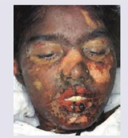
A 28-year-old male presented with a rash on his arms, legs, and face, along with painful ulceration of his lips and mouth, and fever. He developed acute sore throat and conjunctivitis, leading to a diagnosis of acute pharyngitis. Oral penicillin was prescribed. Which of the following can be used in the treatment of the patient's condition, EXCEPT?
What is the primary treatment for Dermatitis herpetiformis?
A patient presents with bullous lesions. What is the characteristic finding on a Tzanck smear?
U-serrated pattern in direct immunofluorescence is seen in:
All are true about Dermatitis herpetiformis EXCEPT:
Intra-epithelial split is seen in which of the following conditions?
A 12-year-old male presented with intensely itchy grouped vesicles on the buttock, trunk, and scalp. On exposure to wheat, they exaggerate. What is the diagnosis?
Granular deposit of IgA at the dermoepidermal junction is seen in which of the following conditions?
A 25-year-old office assistant in a multinational company was taking diclofenac sodium for low back ache for last few weeks. She presents with the following skin lesions. All are true about the image shown except:
Practice by Chapter
Pemphigus Vulgaris
Practice Questions
Pemphigus Foliaceus
Practice Questions
Bullous Pemphigoid
Practice Questions
Cicatricial Pemphigoid
Practice Questions
Dermatitis Herpetiformis
Practice Questions
Epidermolysis Bullosa
Practice Questions
Linear IgA Bullous Dermatosis
Practice Questions
Pemphigoid Gestationis
Practice Questions
Drug-Induced Bullous Disorders
Practice Questions
Immunofluorescence in Bullous Diseases
Practice Questions
Management of Autoimmune Bullous Diseases
Practice Questions
Genetic Counseling in Inherited Blistering Diseases
Practice Questions
Want unlimited practice?
Get full access to all questions, explanations, and performance tracking.
Scan to download app