A patient presents with painful blisters along the chest wall. All of the following tests are useful for diagnosis except:
An elderly patient presents with itchy tense blisters on normal looking skin as well as on urticarial plaques as shown below. The most probable diagnosis is: (AIIMS Nov 2015)

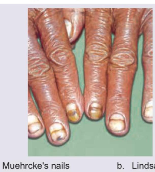
The given picture depicts:

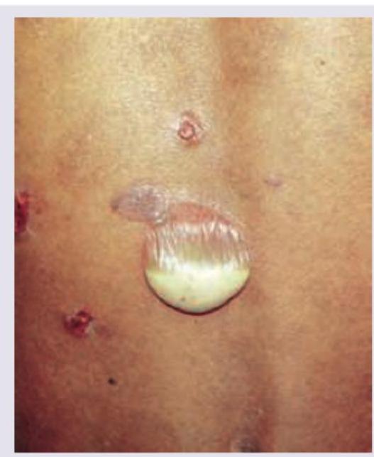
The image shows presence of:

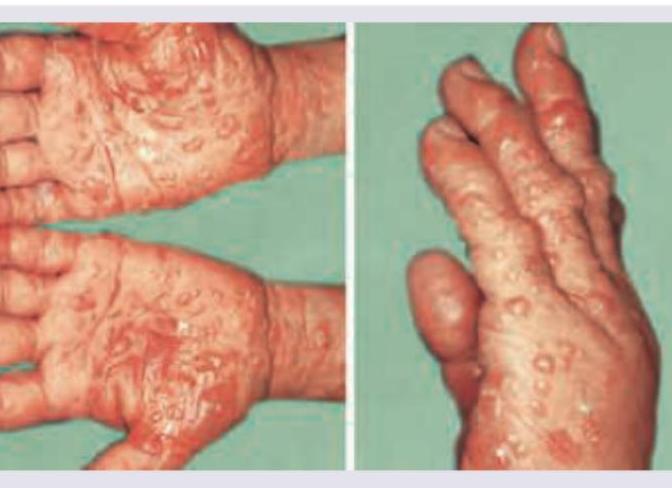
All are true about the lesions shown except:

A patient presents with the skin lesions shown in the image. While evaluating for possible blistering disorders, all of the following conditions could present with similar morphology EXCEPT:

All are true about the lesion shown in the image except:

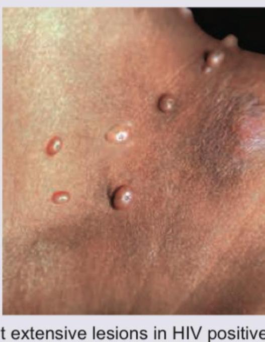
The following image shows a flaccid bulla. This finding is characteristically seen in:

Pseudo Nikolsky sign is seen in all except:
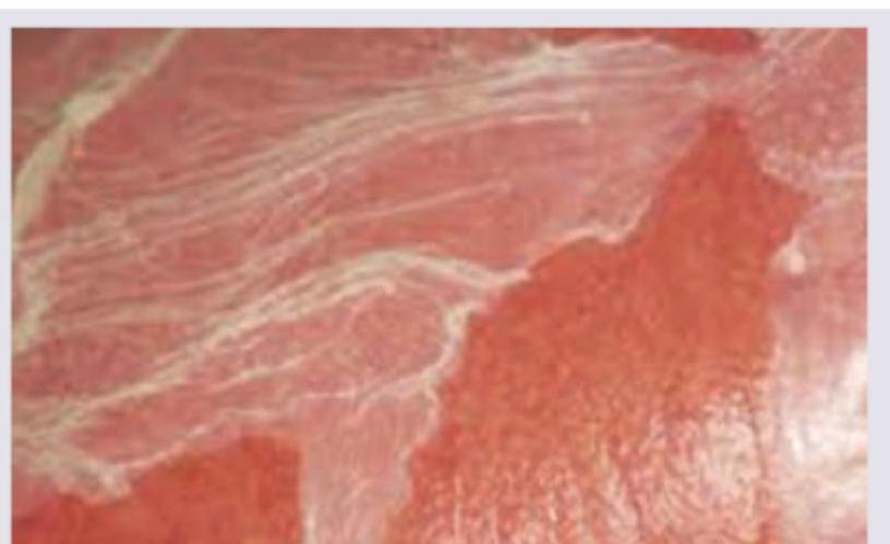
All are true about the lesion shown in a patient with stunting, osmotic diarrhea and anemia except:

Explanation: ***LDH levels*** - **Lactate dehydrogenase (LDH)** is a non-specific enzyme elevated in various conditions causing tissue damage or cellular turnover, but it is not a primary diagnostic tool for herpes zoster. - While systemic inflammation from a severe case of shingles might indirectly affect LDH, it is not used to confirm the diagnosis of this viral infection. *Direct fluorescent antibody* - **Direct fluorescent antibody (DFA)** testing from a blister scraping can rapidly detect varicella-zoster virus (VZV) antigens in cells. - This is a highly sensitive and specific method for confirming VZV infection, differentiating it from other blistering conditions. *Polymerase chain reaction* - **Polymerase chain reaction (PCR)** is a highly sensitive and specific method for detecting VZV DNA from vesicle fluid or crusts. - PCR can rapidly confirm the diagnosis of herpes zoster, especially in atypical presentations or immunocompromised patients. *Tzanck smear* - A **Tzanck smear** involves scraping the base of a blister and examining the cells microscopically for multinucleated giant cells and acantholytic cells. - While not specific for VZV (also seen in HSV infections), it indicates a herpes group viral infection and can be a rapid bedside diagnostic aid.
Explanation: ***Bullous pemphigoid*** - This condition typically presents in **elderly patients** with **itchy, tense blisters** on either normal or erythematous/urticarial skin, which aligns with the clinical description and image. - The blisters in bullous pemphigoid are characterized by **subepidermal blistering**, meaning the epidermis separates from the dermis, resulting in tense, fluid-filled lesions. *Pemphigus vulgaris* - Characterized by **flaccid blisters** that rupture easily, leading to erosions, unlike the tense blisters seen in the image. - Pemphigus vulgaris frequently involves **mucous membranes** and is caused by autoantibodies against desmoglein 1 and 3, leading to intraepidermal blistering (acantholysis). *Linear IgA disease* - This autoimmune blistering condition typically presents with **annular or rosette-shaped lesions** with small peripheral blisters, often referred to as a "string of pearls" appearance, which is not evident in the image. - On **direct immunofluorescence**, it shows a linear deposition of IgA at the dermoepidermal junction. *Dermatitis herpetiformis* - Often presents with **intensely pruritic papules and vesicles** typically found on extensor surfaces (elbows, knees, buttocks), and the lesions are often excoriated due to scratching. - Strongly associated with **celiac disease** and characterized by granular IgA deposits in the dermal papillae on direct immunofluorescence, distinguishing it from the tense blisters seen.
Explanation: ***Tinea imbricata*** - The image shows a distinctive pattern of **concentric, lamellar scales**, often described as "tiles" or "fish scales," which is a hallmark of **tinea imbricata**. - This presentation is caused by specific fungal species, primarily *Trichophyton concentricum*, and is common in certain tropical and subtropical regions. *Toxic epidermal necrolysis* - This condition is characterized by **widespread epidermal detachment** resembling severe burns, leading to large areas of denuded skin and mucosal involvement. - It does not typically present with the **concentric scaly pattern** seen in the image. *Atopic dermatitis* - Characterized by **eczematous lesions**, typically itchy, dry, inflamed skin, often with lichenification in chronic cases. - The appearance is distinct from the **patterned scaling** observed in the image, instead showing erythema, papules, vesicles, and crusting. *Psoriasis* - Psoriasis typically presents as **well-demarcated erythematous plaques** covered with **silvery scales**, often found on extensor surfaces like elbows and knees. - While it involves scaling, it does not exhibit the specific **concentric, imbricated (overlapping) scale pattern** characteristic of the image.
Explanation: ***Muehrcke's nails*** - The image clearly displays characteristic **paired white lines** (leukonychia) separated by normal-appearing nail areas, which are hallmarks of Muehrcke's nails. - These lines are caused by **edema in the nail bed** rather than an abnormality of the nail plate itself, and they do not move with nail growth. *Lindsay nails* - Also known as **"half-and-half" nails**, Lindsay nails feature the **proximal half of the nail plate appearing white** and the **distal half appearing red or pink**, typically associated with chronic kidney disease. - This pattern of discoloration is distinct from the multiple transverse white bands seen in Muehrcke's nails. *Koilonychia* - Koilonychia, or **spoon nails**, describes nails that are **thinned and concave with raised edges**, resembling a spoon. - This condition is often associated with **iron deficiency anemia**, and the appearance in the image does not show this characteristic "spooning." *Beau's lines* - Beau's lines are **transverse depressions or grooves** across the nail plate that occur due to a temporary interruption of nail growth. - They are typically seen as a single, deep furrow that grows out with the nail, unlike the multiple, non-palpable white bands of Muehrcke's nails.
Explanation: ***Potent topical steroids resolve most cases*** - This statement is **false** and is the correct answer to this "EXCEPT" question. - Dyshidrotic eczema (pompholyx) is a **chronic, relapsing condition** that often requires long-term management. - While potent topical steroids are the **mainstay of initial treatment**, they typically provide symptomatic control rather than permanent resolution. - **Recurrences are common**, and many patients require ongoing maintenance therapy or additional treatments such as systemic steroids, immunosuppressants, or phototherapy for severe or refractory cases. *Itchy lesions on palms* - Dyshidrotic eczema is characterized by **intensely pruritic vesicles** that typically appear on the **palms, soles, and lateral aspects of the fingers**. - The image clearly shows vesicular lesions on the palms, consistent with this classic presentation. *Associated with atopic diathesis* - There is a **strong association** between dyshidrotic eczema and **atopic conditions** (atopic dermatitis, allergic rhinitis, asthma). - Approximately **50% of patients** with pompholyx have a personal or family history of atopy. - This atopic association is an important epidemiological feature of the condition. *Painful deep seated vesicles* - The vesicles in dyshidrotic eczema are characteristically **deep-seated** (often described as resembling "tapioca pudding"), especially in the early stages. - They can be both **intensely itchy and painful**, particularly when they are tense and before rupture. - The image demonstrates these characteristic deep vesicular lesions.
Explanation: ***Bullous pemphigoid*** - Presents with **tense bullae** on an erythematous base, typically in elderly patients, unlike the **umbilicated papules** seen in this image. - Involves **subepidermal blistering** with **linear IgG deposition** at the basement membrane zone, not the viral inclusions of Molluscum contagiosum. *Pemphigus vegetans* - A rare variant of pemphigus vulgaris characterized by **vegetating plaques and pustules** in intertriginous areas, not discrete umbilicated lesions. - Shows **intraepidermal acantholysis** with **suprabasal clefting**, histologically distinct from the viral cytopathic changes in Molluscum contagiosum. *Pemphigus vulgaris* - Presents with **flaccid bullae** and painful **mucosal erosions** due to **autoantibodies against desmoglein 1 and 3**. - The **Nikolsky sign** is positive, and lesions are erosive rather than the solid, pearl-like papules characteristic of Molluscum contagiosum. *Pemphigus erythematosus* - Features **erythematous, scaly, crusted lesions** primarily on the **face and upper trunk** with a butterfly distribution. - Combines features of **lupus erythematosus** and pemphigus foliaceus, showing superficial blistering unlike the viral papules in this case.
Explanation: ***Henderson-Patterson bodies are intranuclear bodies*** - **Henderson-Patterson bodies** are characteristic **eosinophilic viral inclusion bodies** found in cells infected with **molluscum contagiosum virus**. - These inclusion bodies are typically found within the **cytoplasm** (intracytoplasmic), not the nucleus. *Giant extensive lesions in HIV positive patients* - Patients with **compromised immune systems**, such as those who are **HIV positive**, can develop unusually large and extensive lesions. - This is due to their inability to mount an effective immune response against the **molluscum contagiosum virus**. *Auto-innoculated lesions* - **Molluscum contagiosum** lesions can spread to other areas of the body through **self-scratching** or contact. - This process, known as **auto-inoculation**, leads to new lesions appearing where the virus made contact with previously unaffected skin. *Needle extirpation followed by trichloro-acetic acid application* - **Needle extirpation** (pricking the central core of the lesion) followed by the application of **trichloroacetic acid (TCA)** is a common treatment method for molluscum contagiosum. - This approach aims to destroy the infected cells and prevent further viral spread.
Explanation: ***Pemphigus vulgaris*** - The image shows a **flaccid bulla** with purulent fluid, characteristic of **pemphigus vulgaris**. This condition is marked by autoantibodies against desmogleins 1 and 3, which are crucial for keratinocyte adhesion, leading to **intraepidermal blistering** and the **Nikolsky sign**. - The flaccid nature of the bulla, often leading to easy rupture and erosions, is a hallmark of superficial blistering in pemphigus vulgaris, caused by the **loss of cell-to-cell adhesion** within the epidermis. *Pemphigus vegetans* - This is a rare variant of pemphigus vulgaris characterized by **vegetating plaques** and **hyperkeratotic lesions**, particularly in intertriginous areas. - While it starts with bullae, the predominant feature is the development of fungating, vegetative lesions rather than the flaccid bulla seen here. *Pemphigus erythematosus* - Pemphigus erythematosus, also known as Senear-Usher syndrome, is considered a localized form of pemphigus foliaceus with features of **lupus erythematosus**. - It presents with **scaling, crusting, and erythematous lesions** resembling lupus, along with superficial bullae, typically on the face and scalp. *Bullous pemphigoid* - Bullous pemphigoid typically presents with **tense bullae** that are less prone to rupture, unlike the flaccid bulla shown in the image. - It is caused by autoantibodies against hemidesmosomal proteins (BP180 and BP230), resulting in **subepidermal blistering**, meaning the blister forms below the epidermis and is therefore more resilient.
Explanation: ***Erythema Multiforme*** - Erythema multiforme is an acute, self-limiting inflammatory dermatosis, and it typically does not present with a **Pseudo Nikolsky sign** as its lesions are usually fixed or have minimal epidermal involvement. - The disease is characterized by target lesions, often in response to infections (e.g., herpes simplex virus) or drugs, with **subepidermal blistering** in some cases but usually without extensive epidermal detachment. *Toxic epidermal Necrolysis* - **Toxic epidermal Necrolysis (TEN)** is a severe mucocutaneous reaction characterized by widespread **epidermal detachment**, making the Nikolsky sign and Pseudo Nikolsky sign positive due to extensive skin fragility and epidermal loss. - This condition involves full-thickness epidermal necrosis, leading to blistering and sloughing of skin that resembles a **severe burn**. *Stevens Johnson Syndrome* - **Stevens-Johnson Syndrome (SJS)** is a less severe form of TEN, but it also features **epidermal detachment** and usually has a positive Nikolsky sign, and therefore also a Pseudo Nikolsky sign. - It involves smaller body surface area detachment compared to TEN but still demonstrates significant epidermal damage and skin fragility. *Staphylococcal scalded skin syndrome* - In **Staphylococcal scalded skin syndrome (SSSS)**, toxins produced by *Staphylococcus aureus* specifically target **desmoglein-1**, causing superficial epidermal splitting and a prominent positive Nikolsky sign (and Pseudo Nikolsky sign). - This condition results in widespread **flaccid blisters** and exfoliation, particularly in children, without full-thickness epidermal necrosis.
Explanation: ***Mucosa is not involved*** - This statement is **FALSE** and is the correct answer to this EXCEPT question. - The clinical presentation with **stunting, osmotic diarrhea, and anemia** clearly indicates **significant intestinal mucosal involvement** and malabsorption. - **Dermatitis herpetiformis** is the cutaneous manifestation of **celiac disease**, which by definition involves an immune-mediated injury to the **small intestinal mucosa** triggered by gluten ingestion. - The gastrointestinal symptoms described are direct evidence of mucosal damage, making this statement incorrect. *IgA antibody against epidermal transglutaminase* - This is TRUE. **Dermatitis herpetiformis** involves **IgA antibodies against epidermal transglutaminase (eTG)**, which deposit in the dermal papillae. - Patients also have IgA antibodies against tissue transglutaminase (tTG) due to the underlying celiac disease. - These antibodies are key diagnostic markers for both conditions. *Intense pruritic lesions* - This is TRUE. **Dermatitis herpetiformis** is classically characterized by **severely pruritic vesicular lesions** with a burning or stinging sensation. - The intense itching often leads to excoriations from scratching before vesicles are even visible. - Lesions typically occur on extensor surfaces (elbows, knees, buttocks, scalp). *Genes encoding DR2 present in all patients* - This statement is also FALSE, but less obviously so. **HLA-DQ2** (90-95%) and **HLA-DQ8** (5-10%) are the primary genetic associations with celiac disease and dermatitis herpetiformis. - If "DR2" refers to HLA-DR2, this is incorrect - DR2 is associated with other autoimmune conditions like multiple sclerosis, not celiac disease. - Even if this were meant to say DQ2, it would still be false as not 100% of patients carry these genes.
Pemphigus Vulgaris
Practice Questions
Pemphigus Foliaceus
Practice Questions
Bullous Pemphigoid
Practice Questions
Cicatricial Pemphigoid
Practice Questions
Dermatitis Herpetiformis
Practice Questions
Epidermolysis Bullosa
Practice Questions
Linear IgA Bullous Dermatosis
Practice Questions
Pemphigoid Gestationis
Practice Questions
Drug-Induced Bullous Disorders
Practice Questions
Immunofluorescence in Bullous Diseases
Practice Questions
Management of Autoimmune Bullous Diseases
Practice Questions
Genetic Counseling in Inherited Blistering Diseases
Practice Questions
Get full access to all questions, explanations, and performance tracking.
Start For Free