U-serrated pattern in direct immunofluorescence is seen in:
Acantholysis is characteristic of which of the following conditions?
In pemphigus, circulating antibodies attack which components?
All are true about Dermatitis herpetiformis EXCEPT:
Which of the following antigens are associated with cicatricial pemphigoid?
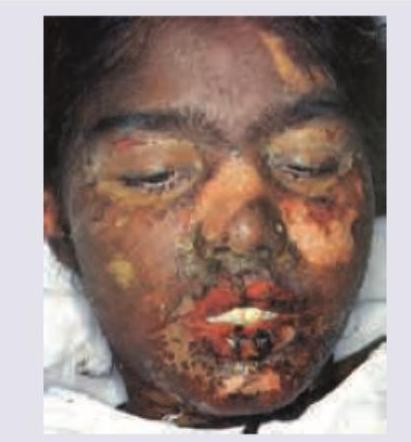
What percentage of skin involvement is characteristic of toxic epidermal necrolysis?
Intra-epithelial split is seen in which of the following conditions?
A 12-year-old male presented with intensely itchy grouped vesicles on the buttock, trunk, and scalp. On exposure to wheat, they exaggerate. What is the diagnosis?
Desmoplakin is the target antigen in which of the following conditions?
A 25-year-old office assistant in a multinational company was taking diclofenac sodium for low back ache for last few weeks. She presents with the following skin lesions. All are true about the image shown except:

Explanation: ### Explanation The correct answer is **Epidermolysis bullosa acquisita (EBA)**. **1. Why EBA is correct:** Direct Immunofluorescence (DIF) of perilesional skin in subepidermal autoimmune bullous diseases typically shows linear IgG and C3 deposits along the basement membrane zone (BMZ). However, using **Salt-Split Skin (SSS)** or high-resolution DIF, two distinct patterns emerge: * **U-serrated pattern:** Characteristic of **EBA**. The immune deposits occur below the lamina densa (targeting Type VII collagen). These deposits follow the contours of the dermal papillae, resembling the letter "U" or "grass-like" arcs. * **N-serrated pattern:** Characteristic of **Bullous Pemphigoid**. The deposits occur in the lamina lucida (targeting BP180/230), appearing as "saw-tooth" or "N" shapes. **2. Why other options are incorrect:** * **Bullous pemphigoid:** Shows an **N-serrated pattern** on DIF. On Salt-split skin, the fluorescence is on the **roof** (epidermal side). * **Linear IgA disease:** Characterized by linear deposits of **IgA** (not IgG) along the BMZ. It does not typically show the U-serrated morphology. * **Dermatitis herpetiformis:** Shows **granular IgA deposits** at the tips of dermal papillae, not a linear serrated pattern. **3. High-Yield Clinical Pearls for NEET-PG:** * **Target Antigen in EBA:** Type VII Collagen (anchoring fibrils). * **Salt-Split Skin Test:** In EBA, the fluorescence is on the **floor** (dermal side) of the split. In Bullous Pemphigoid, it is on the **roof** (epidermal side). * **Clinical variants of EBA:** Mechanobullous (classic) and Inflammatory (resembling BP). * **Mnemonic:** **E**BA = **B**ottom (Floor) / **U**-serrated. **B**P = **T**op (Roof) / **N**-serrated.
Explanation: **Explanation:** **Acantholysis** is the hallmark pathological process of the **Pemphigus** group of diseases. It refers to the loss of intercellular connections (desmosomes) between keratinocytes, leading to the formation of intraepidermal clefts and blisters. In **Pemphigus vulgaris**, IgG autoantibodies target **Desmoglein 3** (and sometimes Desmoglein 1), resulting in "row of tombstone" appearance on histology due to the loss of cohesion between suprabasal cells. **Why other options are incorrect:** * **Pemphigoid (Bullous Pemphigoid):** This is a **subepidermal** blistering disease. The pathology involves autoantibodies against BP180 and BP230 in the hemidesmosomes. There is no acantholysis; instead, the entire epidermis detaches from the dermis. * **Erythema Multiforme:** This is a hypersensitivity reaction characterized by "target lesions." Histologically, it shows vacuolar degeneration of the basal layer and individual keratinocyte necrosis, not acantholysis. * **Dermatitis Herpetiformis:** This is an IgA-mediated autoimmune disease associated with Celiac disease. It is characterized by **subepidermal** blisters and neutrophilic microabscesses at the dermal papillary tips. **High-Yield Clinical Pearls for NEET-PG:** * **Nikolsky Sign:** Positive in Pemphigus (due to acantholysis) but negative in Pemphigoid. * **Tzanck Smear:** Shows **Acantholytic cells (Tzanck cells)**—large, round keratinocytes with hyperchromatic nuclei and a peripheral halo of cytoplasm. * **Immunofluorescence:** Pemphigus shows a **"fishnet" or "lace-like"** pattern of IgG deposition, whereas Bullous Pemphigoid shows **linear** IgG/C3 deposition along the basement membrane zone.
Explanation: **Explanation:** Pemphigus is a group of life-threatening autoimmune blistering diseases characterized by **acantholysis** (loss of cell-to-cell adhesion). The underlying pathophysiology involves the production of IgG autoantibodies against **Desmogleins (Dsg)**, which are calcium-dependent transmembrane glycoproteins belonging to the cadherin family. These proteins are vital components of **desmosomes**, the "glue" that holds keratinocytes together. * **Pemphigus Vulgaris (PV):** The most common form. Antibodies primarily target **Dsg 3** (found in mucosal surfaces) and **Dsg 1** (found in the skin). This leads to mucosal ulcers and flaccid skin bullae. * **Pemphigus Foliaceus (PF):** Antibodies target **Dsg 1** only, resulting in superficial blisters and crusting without mucosal involvement. **Analysis of Options:** * **Option B (Correct):** Dsg 1 and Dsg 3 are the primary targets in the Pemphigus group. * **Options A, C, and D (Incorrect):** While Desmoglein 2 and 4 exist, they are not the primary antigenic targets in classic Pemphigus. Dsg 2 is expressed in simple epithelia and the heart, while Dsg 4 is primarily expressed in hair follicles. **High-Yield Clinical Pearls for NEET-PG:** 1. **Nikolsky Sign:** Positive (gentle pressure on normal-looking skin causes perilesional skin to peel). 2. **Tzanck Smear:** Shows **Acantholytic cells** (Tzanck cells/Rowley cells)—large, round keratinocytes with hyperchromatic nuclei. 3. **Histopathology:** Characterized by **suprabasal splitting** and a "row of tombstones" appearance (basal layer remains attached to the basement membrane). 4. **Direct Immunofluorescence (DIF):** Shows a characteristic **"fishnet" or "lace-like"** pattern of IgG and C3 deposits in the intercellular spaces.
Explanation: ### Explanation **Dermatitis Herpetiformis (DH)** is a chronic, autoimmune blistering disease characterized by intensely pruritic, grouped vesicles. The correct answer is **B** because DH is a **subepidermal** blistering disease, not epidermal. #### 1. Why Option B is the Correct Answer (The Exception) In DH, the primary pathology occurs at the **dermo-epidermal junction**. Neutrophils accumulate at the tips of the dermal papillae (forming **microabscesses**), leading to the separation of the epidermis from the dermis. This results in **subepidermal bullae**. Intraepidermal or "epidermal" bullae are characteristic of the Pemphigus group of diseases, not DH. #### 2. Analysis of Other Options * **Option A:** DH is notoriously known as "the itch that rashes." The pruritus is so intense that patients often present with excoriations and crusts rather than intact vesicles. * **Option C:** The gold standard for diagnosis is Direct Immunofluorescence (DIF) of perilesional skin, which shows **granular IgA deposits** localized in the **papillary tips**. * **Option D:** DH is considered the cutaneous manifestation of **Celiac disease**. Nearly 90% of patients have associated gluten-sensitive enteropathy (though it may be asymptomatic), and both conditions share the **HLA-DQ2/DQ8** haplotype. #### 3. High-Yield Clinical Pearls for NEET-PG * **Distribution:** Symmetrical involvement of extensors (elbows, knees, buttocks, and scalp). * **Histopathology:** Neutrophilic microabscesses at the dermal papillary tips (Pierard’s microabscesses). * **Treatment of Choice:** **Dapsone** (provides rapid relief from itching) along with a strict **Gluten-Free Diet (GFD)**. * **Target Antigen:** Epidermal Transglutaminase (eTG/TG3).
Explanation: **Explanation:** **Cicatricial Pemphigoid (Mucous Membrane Pemphigoid)** is a chronic, autoimmune subepidermal blistering disease primarily affecting the mucous membranes (oral, ocular, and genital) and frequently leading to scarring (cicatrization). 1. **Why Option A is Correct:** The pathogenesis involves autoantibodies directed against components of the dermo-epidermal junction. The most common target antigens are **BPAG2 (BP180)**, specifically the C-terminus, and **Laminin 332 (Epiligrin)**. * **BPAG2** is associated with the mucosal-predominant form. * **Epiligrin (Laminin 332)** is a high-yield association because patients with anti-epiligrin cicatricial pemphigoid have a significantly **increased risk of solid organ malignancies** (adenocarcinomas). 2. **Why Other Options are Incorrect:** * **Options B, C, and D:** These refer to Human Leukocyte Antigen (HLA) types. While certain HLAs (like HLA-DQB1*0301) are associated with Cicatricial Pemphigoid, the question specifically asks for **antigens** (the proteins targeted by antibodies), not genetic susceptibility markers. HLA DR5, B8, and DR3 are more commonly linked to other autoimmune conditions like Dermatitis Herpetiformis or Systemic Lupus Erythematosus. **High-Yield Clinical Pearls for NEET-PG:** * **Site:** Most common site is the **oral mucosa** (desquamative gingivitis), but the most serious complication is **ocular involvement** (symblepharon, ankyloblepharon, and blindness). * **Direct Immunofluorescence (DIF):** Shows linear IgG and C3 deposits at the basement membrane zone. * **Salt-split skin test:** Fluorescence occurs on the **roof** (epidermal side) if targeting BP180, or the **floor** (dermal side) if targeting Laminin 332. * **Key Association:** Always screen for internal malignancy if anti-epiligrin antibodies are present.
Explanation: Toxic Epidermal Necrolysis (TEN) and Stevens-Johnson Syndrome (SJS) are severe mucocutaneous adverse drug reactions characterized by extensive keratinocyte apoptosis and epidermal detachment. They are classified as a spectrum based on the percentage of **Total Body Surface Area (TBSA)** affected by skin detachment. ### **Explanation of Options:** * **Option D (Correct):** **Greater than 30%** involvement is the diagnostic threshold for **Toxic Epidermal Necrolysis (TEN)**. This represents the most severe end of the spectrum, often associated with high mortality and multi-organ involvement. * **Option A:** **Less than 10%** involvement is characteristic of **Stevens-Johnson Syndrome (SJS)**. * **Options B & C:** The range of **10% to 30%** involvement is classified as **SJS/TEN Overlap**. ### **High-Yield NEET-PG Clinical Pearls:** 1. **Nikolsky Sign:** This is characteristically **positive** in SJS/TEN (slight pressure on the skin causes exfoliation of the outermost layer). 2. **Etiology:** Most commonly triggered by drugs like **Sulfonamides**, **Antiepileptics** (Phenytoin, Carbamazepine), **Allopurinol**, and **NSAIDs**. 3. **Histopathology:** Shows **full-thickness epidermal necrosis** with a subepidermal split and minimal dermal inflammation. 4. **SCORTEN:** A validated scoring system used to predict the prognosis and mortality risk in TEN patients. 5. **Management:** Immediate withdrawal of the offending drug and supportive care in a **Burn Unit** or ICU is the gold standard.
Explanation: **Explanation:** The level of split in blistering diseases is a high-yield concept for NEET-PG. Blisters are classified based on whether the cleavage occurs within the epidermis (**Intra-epidermal/Intra-epithelial**) or below it (**Sub-epidermal**). **1. Why Pemphigus Vulgaris is correct:** Pemphigus vulgaris is an autoimmune disease characterized by IgG antibodies against **Desmoglein 3** (and 1). These proteins are components of desmosomes, which hold keratinocytes together. The destruction of these "bridges" leads to **acantholysis** (loss of intercellular adhesion), resulting in an **intra-epithelial split**. Specifically, in Pemphigus Vulgaris, the split occurs just above the basal layer, creating a "tombstone appearance." **2. Why other options are incorrect:** * **Bullous Pemphigoid:** This is a **sub-epidermal** blistering disease. Autoantibodies (anti-BP180 and BP230) target the hemidesmosomes at the dermo-epidermal junction, causing the entire epidermis to lift off the dermis. * **Epidermolysis Bullosa (EB):** While EB Simplex is intra-epidermal, the term "Epidermolysis Bullosa" generally encompasses a group of genetic disorders where the majority of types (Junctional and Dystrophic) involve **sub-epidermal** cleavage. In the context of standard MCQ patterns, Pemphigus is the classic prototype for intra-epithelial splits. **Clinical Pearls for NEET-PG:** * **Nikolsky Sign:** Positive in Pemphigus (intra-epidermal) and negative in Bullous Pemphigoid (sub-epidermal). * **Tzanck Smear:** Shows **Acantholytic cells** (Tzanck cells) in Pemphigus. * **DIF Pattern:** Pemphigus shows a **"Fish-net"** or "Lace-like" pattern; Bullous Pemphigoid shows a **Linear** pattern along the basement membrane zone.
Explanation: ### Explanation **Correct Answer: B. Dermatitis herpetiformis** **Concept:** Dermatitis herpetiformis (DH) is a chronic, autoimmune blistering disease characterized by intensely pruritic, symmetric, grouped vesicles (herpetiform) typically involving the extensor surfaces (elbows, knees), buttocks, and scalp. The hallmark of DH is its strong association with **Gluten-Sensitive Enteropathy (Celiac Disease)**. The ingestion of wheat (gluten) triggers the production of IgA antibodies against tissue transglutaminase (tTG), which cross-react with epidermal transglutaminase (eTG), leading to subepidermal blister formation. **Why the other options are incorrect:** * **Herpes simplex:** While it presents with grouped vesicles, it is typically localized (e.g., labial or genital), acute, and not triggered by dietary wheat. * **Pemphigus vulgaris:** This is an intraepidermal blistering disease characterized by flaccid bullae and painful oral erosions. It is not intensely itchy and lacks the dietary association with gluten. * **Bullous impetigo:** A superficial bacterial infection (usually *S. aureus*) presenting with fragile bullae that leave a "collarette" of scale. It is common in children but is not chronic or triggered by wheat. **High-Yield Clinical Pearls for NEET-PG:** * **Gold Standard Diagnosis:** Direct Immunofluorescence (DIF) showing **granular IgA deposits** at the tips of dermal papillae. * **Histopathology:** Characterized by **neutrophilic microabscesses** (Papillary tip abscesses) and subepidermal clefting. * **Treatment of Choice:** **Dapsone** (provides rapid relief of itch) along with a strict **Gluten-Free Diet**. * **Associated HLA:** Strongly associated with **HLA-DQ2** and **HLA-DQ8**.
Explanation: ### Explanation **Correct Answer: B. Paraneoplastic pemphigus** **Medical Concept:** Paraneoplastic Pemphigus (PNP) is a severe autoimmune blistering disease associated with underlying malignancies (most commonly Non-Hodgkin Lymphoma and CLL). Unlike other forms of pemphigus, PNP is characterized by a **polymorphic** autoantibody profile. The autoantibodies target not only the desmogleins (Dsg 1 and 3) but also members of the **Plakin family**, specifically **Desmoplakin I and II**, Bullous Pemphigoid Antigen 1 (BP230), Periplakin, and Envoplakin. These proteins are intracellular components of the desmosome and hemidesmosome, and their destruction leads to extensive acantholysis. **Analysis of Incorrect Options:** * **A. Pemphigus vulgaris:** The primary target antigens are **Desmoglein 3** (mucosal-dominant) and **Desmoglein 1** (mucocutaneous). It does not typically involve plakin proteins. * **C. Drug-induced pemphigus:** This most commonly mimics Pemphigus foliaceus or vulgaris. The antigens are usually **Desmoglein 1 or 3**. Common culprits include Thiol group drugs like D-penicillamine and Captopril. * **D. Pemphigus foliaceous:** The target antigen is strictly **Desmoglein 1**, leading to very superficial (subcorneal) blisters. **High-Yield Clinical Pearls for NEET-PG:** * **Most common association:** Non-Hodgkin Lymphoma (NHL) is the most frequent malignancy associated with PNP. In children, it is often associated with Castleman disease. * **Clinical Hallmark:** Severe, intractable **stomatitis** (painful oral ulcerations) that involves the tongue and extends to the oropharynx. * **Unique Feature:** PNP can involve the respiratory epithelium, leading to **Bronchiolitis Obliterans**, which is a major cause of mortality. * **Immunofluorescence:** Direct Immunofluorescence (DIF) shows IgG and C3 in the intercellular spaces (fishnet pattern) AND along the basement membrane zone (linear pattern).
Explanation: ***>30% BSA involvement in Stevens-Johnson syndrome*** - Stevens-Johnson syndrome (SJS) is defined by skin detachment affecting **less than 10%** of the body surface area (BSA). - Involvement of **>30% BSA** is characteristic of **Toxic Epidermal Necrolysis (TEN)**, which is a more severe form on the SJS-TEN spectrum. - Therefore, this statement is **FALSE** for SJS and is the correct answer to this "except" question. *Hemorrhagic crusting of lips* - The image shows extensive **hemorrhagic crusting and erosions** on the lips, which is a hallmark feature of SJS/TEN. - This mucosal involvement causes significant pain, difficulty eating, and increased infection risk. - This statement is **TRUE** about the condition shown. *Apoptotic keratinocytes* - Histopathologically, SJS and TEN are characterized by widespread **keratinocyte apoptosis**, leading to full-thickness epidermal necrosis and detachment. - This programmed cell death is the key pathophysiologic mechanism underlying the severe skin damage. - This statement is **TRUE** about SJS/TEN. *Systemic involvement leads to lung and kidney malfunction* - SJS/TEN can cause **severe systemic complications** affecting multiple organs including lungs (pneumonitis, ARDS), kidneys (acute tubular necrosis), liver, and GI tract. - Systemic complications and secondary infections are major causes of morbidity and mortality. - This statement is **TRUE** about severe SJS/TEN cases.
Pemphigus Vulgaris
Practice Questions
Pemphigus Foliaceus
Practice Questions
Bullous Pemphigoid
Practice Questions
Cicatricial Pemphigoid
Practice Questions
Dermatitis Herpetiformis
Practice Questions
Epidermolysis Bullosa
Practice Questions
Linear IgA Bullous Dermatosis
Practice Questions
Pemphigoid Gestationis
Practice Questions
Drug-Induced Bullous Disorders
Practice Questions
Immunofluorescence in Bullous Diseases
Practice Questions
Management of Autoimmune Bullous Diseases
Practice Questions
Genetic Counseling in Inherited Blistering Diseases
Practice Questions
Get full access to all questions, explanations, and performance tracking.
Start For Free