What is the diagnosis of an umbilicated, pearly white, asymptomatic skin lesion?
An 18-year-old man has facial and upper back lesions that have waxed and waned for the past 6 years. On physical examination, there are 0.3- to 0.9-cm comedones, erythematous papules, nodules, and pustules most numerous on the lower face and posterior upper trunk. Other family members have been affected by this condition at a similar age. The lesions worsen during a 5-day cruise to the Adriatic. Which of the following organisms is most likely to play a key role in the pathogenesis of these lesions?
A patient, a resident of Himachal Pradesh, presented with a series of ulcers in a row on his right leg. The biopsy from the affected area was taken and cultured on Sabouraud's dextrose agar. What is the most likely causative organism?
A young child of 7 years of age is seen with indurated ulcers, lymphadenopathy and fever. The likely treatment is:
Statement 1 - A 59-year-old patient presents with flaccid bullae. Histopathology shows a suprabasal acantholytic split. Statement 2 - The row of tombstones appearance is diagnostic of Pemphigus vulgaris.
Which of the following conditions is caused by Staphylococcus aureus?
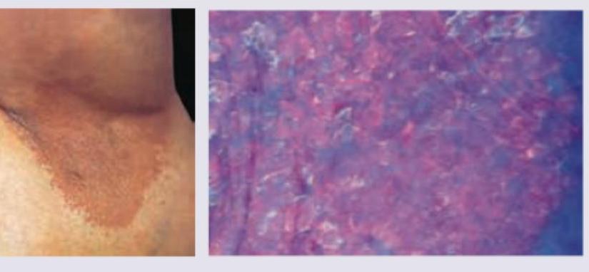
A 35-year-old obese woman presents with recurrent lesions in both axilla in summer season. Wood lamp examination is shown. The diagnosis is:

What is the treatment for granuloma inguinale?
A patient presents with multiple hypopigmented and hypesthetic patches on the lateral aspect of the forearm, with abundant acid-fast bacilli (AFB) and granulomatous inflammation on histology. What is the most likely diagnosis?
Erysipelas is caused by which bacterium?
Explanation: ***Molluscum contagiosum*** - This **viral skin infection** typically presents with **multiple, small (2-5 mm), firm, pearly, dome-shaped papules** that have a **central umbilication**. - The lesions are usually **asymptomatic**, as described, though they can occasionally be itchy or inflamed. - Caused by a **poxvirus** and is highly contagious through direct contact. *EBV* - **Epstein-Barr Virus (EBV)** is primarily associated with **infectious mononucleosis**, which presents with fever, sore throat, and lymphadenopathy, not umbilicated skin lesions. - EBV can cause oral hairy leukoplakia in immunocompromised individuals, which is a white lesion, but it is **not pearly, umbilicated, or dome-shaped**. *HSV* - **Herpes Simplex Virus (HSV)** causes lesions that are typically **grouped vesicles on an erythematous base** that evolve into erosions or ulcers. - HSV lesions are often **painful or itchy** and **do not appear as pearly, umbilicated papules**. *None of the options* - This is incorrect because **Molluscum contagiosum** perfectly matches the clinical description of umbilicated, pearly white, asymptomatic skin lesions. - The classic **central umbilication** is the pathognomonic feature that distinguishes molluscum from other viral skin infections.
Explanation: ***Propionibacterium acnes*** (now *Cutibacterium acnes*) - The presence of **comedones, papules, nodules, and pustules** on the face and upper back in an 18-year-old is classic for **acne vulgaris**. - **_P. acnes_** is a commensal bacterium that proliferates in clogged hair follicles, contributing to inflammation and lesion formation in acne due to its lipolytic activity and immune-activating properties. *Herpes simplex virus type 1* - **HSV-1** typically causes **oral herpes (cold sores)** or **genital herpes**, characterized by painful vesicles and ulcers. - The described lesions (comedones, papules, nodules, pustules) are not characteristic of HSV-1 infection. *Group A β-hemolytic streptococcus* - **Group A Strep** causes infections like **pharyngitis (strep throat)**, **impetigo**, or **cellulitis**, which are typically acute and rapidly spreading. - Its presence is not associated with chronic, polymorphic lesions characteristic of acne. *Mycobacterium leprae* - **_M. leprae_** is the causative agent of **leprosy**, presenting with skin lesions, nerve damage, and other systemic effects. - The skin lesions of leprosy are typically macules, papules, or nodules with sensory loss, not the comedones and pustules seen in acne.
Explanation: ***Sporothrix schenckii*** - The presentation of "ulcers in a row" on the leg is highly suggestive of **lymphocutaneous sporotrichosis**, a characteristic finding where the infection spreads via lymphatic drainage. - This fungus is endemic in certain regions including parts of **Himachal Pradesh**, and is typically acquired through contact with contaminated soil or plant material (e.g., rose thorns, sphagnum moss). - Grows well on **Sabouraud's dextrose agar**, producing characteristic colonies. *Cladosporium spp.* - While *Cladosporium* can cause **phaeohyphomycosis** or allergic fungal sinusitis, it does not typically present with the classic lymphocutaneous lesions described. - These fungi are common environmental contaminants and their infections are usually associated with chronic skin lesions, not a linear spread of ulcers. *Pseudoallescheria boydii* - *Pseudoallescheria boydii* is a common cause of **mycetoma** (Madura foot), characterized by chronic, destructive lesions with granulomas and sinus tracts that discharge grains. - This presentation is distinct from the linear ulcerative lesions described in the patient. *Nocardia brasiliensis* - *Nocardia brasiliensis* is a bacterium (an actinomycete) that causes **actinomycetoma**, characterized by chronic, suppurative lesions with sinus tracts discharging grains. - The characteristic **"ulcers in a row"** (lymphocutaneous spread pattern) is **not typical** of Nocardia infection, which presents as localized mycetoma rather than ascending lymphatic involvement. - While Nocardia can grow on some fungal media, the clinical presentation is the key distinguishing feature here.
Explanation: ***Systemic antibiotics*** - This clinical triad of **indurated ulcers, lymphadenopathy, and fever** in a child is highly suggestive of **ulceroglandular tularemia** (Francisella tularensis), **cat-scratch disease** (Bartonella henselae), or **atypical mycobacterial infection**. - **Tularemia** presents with a painful ulcer at the inoculation site with regional lymphadenopathy and systemic symptoms - treated with **streptomycin or gentamicin**. - **Cat-scratch disease** may present similarly after feline contact - treated with **azithromycin**. - **Atypical mycobacteria** (M. marinum) cause "swimming pool granuloma" with similar features - requiring **clarithromycin and rifampicin**. - **Systemic antibiotic therapy is essential** to prevent complications and disease progression. *Symptomatic treatment* - **Symptomatic treatment alone is inadequate** for bacterial infections presenting with indurated ulcers and lymphadenopathy. - While fever and pain management may be adjunctive, **definitive antimicrobial therapy is required** for these infectious conditions. - Failure to treat appropriately can lead to **systemic dissemination** and serious complications. *Excise the lesion* - **Surgical excision is not the primary treatment** for infectious ulcers with lymphadenopathy. - Excision may be considered for **localized atypical mycobacterial lymphadenitis** that fails medical therapy, but is not first-line. - The presence of **systemic symptoms (fever)** indicates need for medical rather than surgical management. *I.V. fluids* - **Intravenous fluids are supportive therapy** for dehydration, not definitive treatment. - The clinical presentation requires **antimicrobial therapy**, not just hydration. - IV fluids may be needed as adjunctive therapy if the child is unable to maintain oral hydration, but do not address the underlying infection.
Explanation: ***Correct: Statements 1 & 2 are correct, 2 is not explaining 1*** **Analysis of Statement 1:** - A 59-year-old patient with **flaccid bullae** and **suprabasal acantholytic split** on histopathology is the classic presentation of **Pemphigus vulgaris** - The flaccid (easily ruptured) nature of bullae distinguishes it from tense bullae seen in bullous pemphigoid - The suprabasal location of the split (just above the basal layer) with acantholysis (loss of cell-to-cell adhesion) is pathognomonic - **Statement 1 is CORRECT** ✓ **Analysis of Statement 2:** - The **"row of tombstones" or "tombstone appearance"** is indeed a diagnostic histopathological feature of Pemphigus vulgaris - This appearance results from basal keratinocytes remaining attached to the basement membrane while suprabasal cells separate due to acantholysis - The intact basal cells standing upright resemble a row of tombstones - **Statement 2 is CORRECT** ✓ **Does Statement 2 explain Statement 1?** - Statement 2 describes a **histopathological appearance** (tombstone pattern) that is a **consequence** of the suprabasal split - However, it does NOT explain the **underlying cause** of the flaccid bullae or the suprabasal split - The true explanation involves **IgG autoantibodies against desmoglein 3 (and desmoglein 1)**, which attack intercellular adhesion structures (desmosomes), causing **acantholysis** - Therefore, **Statement 2 does NOT explain Statement 1** ✗ *Incorrect: Statement 2 is the correct explanation for Statement 1* - While both statements describe features of Pemphigus vulgaris, the tombstone appearance is a descriptive finding, not an explanatory mechanism *Incorrect: Statements 1 and 2 are incorrect* - Both statements are medically accurate descriptions of Pemphigus vulgaris features *Incorrect: Statement 1 is incorrect* - Statement 1 correctly describes the cardinal clinical and histopathological features of Pemphigus vulgaris
Explanation: ***Bullous impetigo*** - Bullous impetigo is a superficial skin infection characterized by **blisters (bullae)**, and is specifically caused by **Staphylococcus aureus** producing exfoliative toxins. - The toxins produced by *S. aureus* cause intraepidermal cleavage, leading to the formation of the characteristic **flaccid bullae**. *Corynebacterium minutissimum infection* - *Corynebacterium minutissimum* causes **erythrasma**, a chronic superficial skin infection characterized by well-demarcated reddish-brown patches, often in intertriginous areas. - It does not cause bullous impetigo and is typically diagnosed by its coral-red fluorescence under a **Wood's lamp**. *Haemophilus ducreyi infection* - *Haemophilus ducreyi* is the causative agent of **chancroid**, a sexually transmitted infection characterized by painful genital ulcers with a necrotic base and often accompanied by swollen, tender regional lymph nodes. - It is not associated with skin blistering or bullous impetigo. *Propionibacterium acnes infection* - *Propionibacterium acnes* (now *Cutibacterium acnes*) is a bacterium commonly implicated in **acne vulgaris**, contributing to inflammation and comedone formation within hair follicles. - It causes inflammatory lesions like papules, pustules, nodules, and cysts, rather than bullous lesions.
Explanation: ***Erythrasma*** - Erythrasma is a superficial bacterial infection caused by **Corynebacterium minutissimum**, which commonly presents as red-brown patches in intertriginous areas like the axilla, especially in obese individuals and warm, humid conditions (summer season). - The distinctive **coral-red fluorescence under Wood's lamp** is due to porphyrin production by the bacteria, which is a classic diagnostic feature of erythrasma, as shown in the image. *Ecthyma* - Ecthyma is a deeper form of impetigo characterized by **ulcerative lesions with a thick, adherent crust** that extend into the dermis. - It is typically caused by *Streptococcus pyogenes* and sometimes *Staphylococcus aureus*, and would not exhibit coral-red fluorescence under Wood's lamp. *Impetigo contagiosa* - Impetigo contagiosa (non-bullous impetigo) presents with **honey-colored crusted lesions**, usually on the face and extremities. - While also a bacterial skin infection, it is typically caused by *Staphylococcus aureus* or *Streptococcus pyogenes* and does not show coral-red fluorescence under Wood's lamp. *Bullous impetigo* - Bullous impetigo is characterized by **flaccid bullae** (blisters) that rupture to form thin, varnish-like crusts, primarily caused by *Staphylococcus aureus* producing exfoliative toxins. - Similar to other forms of impetigo, it does not produce the coral-red fluorescence under Wood's lamp.
Explanation: ***Azithromycin*** - **Azithromycin** is the recommended first-line treatment for **granuloma inguinale** (donovanosis) caused by *Klebsiella granulomatis*. - Current recommended regimens: **Azithromycin 1g orally once weekly** OR **500mg daily for at least 3 weeks** (until all lesions have completely healed). - Preferred due to excellent tissue penetration, good efficacy, and convenient dosing that improves patient compliance. *Tetracycline* - **Tetracycline** (500mg four times daily) was historically used but has been largely replaced by **doxycycline** (100mg twice daily) as the preferred tetracycline-class antibiotic. - While effective against *Klebsiella granulomatis*, it requires frequent dosing leading to poor adherence. - **Doxycycline** (not listed here) is actually considered a co-first-line option alongside azithromycin in current CDC guidelines. *Clarithromycin* - **Clarithromycin** is a macrolide antibiotic but is not a recommended first-line agent for granuloma inguinale. - Limited clinical data supports its use for this condition, and it is not included in standard treatment guidelines. - Azithromycin from the same macrolide class is preferred due to better-established efficacy. *Streptomycin* - **Streptomycin** is an aminoglycoside antibiotic primarily used for mycobacterial infections (e.g., tuberculosis, plague). - Not indicated for granuloma inguinale as *Klebsiella granulomatis* responds well to macrolides (azithromycin) and tetracyclines (doxycycline). - Requires parenteral administration and has significant toxicity concerns (ototoxicity, nephrotoxicity).
Explanation: ### Explanation The correct answer is **Borderline leprosy (C)**. #### 1. Why Borderline Leprosy is Correct The diagnosis of leprosy is based on the Ridley-Jopling classification, which correlates clinical features with the host's immune response. * **Clinical Presentation:** The presence of multiple hypopigmented and **hypesthetic** (reduced sensation) patches is characteristic of the borderline spectrum. * **Histopathology:** The mention of **abundant acid-fast bacilli (AFB)** alongside **granulomatous inflammation** is the key differentiator. In the borderline spectrum (specifically Borderline Lepromatous - BL), the cell-mediated immunity is low enough to allow significant bacillary multiplication (high Bacterial Index), yet high enough to still form organized granulomas. #### 2. Why Other Options are Incorrect * **A. Tuberculoid leprosy (TT):** Characterized by high immunity. Clinically, there are very few lesions (1-3) with complete anesthesia. Histologically, granulomas are well-formed, but **AFB are absent** (paucibacillary). * **B. Intermediate leprosy:** This is an early, transitory stage. It usually presents as a single, ill-defined macule with vague sensory loss. It does not show abundant AFB or well-developed granulomatous inflammation. * **D. Lepromatous leprosy (LL):** Characterized by negligible immunity. While AFB are extremely abundant (globi), the histology shows **diffuse histiocytic infiltration** (Virchow cells/foam cells) rather than organized granulomatous inflammation. #### 3. NEET-PG High-Yield Pearls * **Pathognomonic sign:** Asymmetrical nerve enlargement is typical of Borderline Leprosy. * **Bacterial Index (BI):** TT (0), BT (0-1+), BB (3-4+), BL (4-5+), LL (5-6+). * **Lepromin Test:** Strongly positive in TT, negative in LL. It measures delayed-type hypersensitivity (prognostic, not diagnostic). * **Treatment:** WHO MDT for Multibacillary (MB) leprosy (including Borderline and LL) lasts 12 months, whereas Paucibacillary (PB) lasts 6 months.
Explanation: ### Explanation **Correct Answer: C. Streptococcus pyogenes** **Medical Concept:** Erysipelas is a distinct clinical variant of superficial cellulitis. It is primarily caused by **Group A Beta-hemolytic Streptococci (GABHS)**, most commonly ***Streptococcus pyogenes***. The infection involves the upper dermis and superficial lymphatics. Characteristically, it presents as a well-demarcated, fiery-red, edematous, and tender plaque. The "sharp borders" are a hallmark feature because the infection is superficial, allowing for a clear distinction between involved and uninvolved skin. **Analysis of Incorrect Options:** * **A. *Staphylococcus aureus*:** While *S. aureus* is the most common cause of **Cellulitis** (which involves the deeper dermis and subcutaneous fat), it is rarely the primary cause of classic Erysipelas. *S. aureus* is more associated with purulent infections like furuncles and abscesses. * **B. *Staphylococcus albus*:** Now known as *Staphylococcus epidermidis*, this is a commensal organism of the skin flora and is generally non-pathogenic unless it involves prosthetic implants or biofilms. * **C. *Haemophilus*:** *Haemophilus influenzae* was historically a common cause of facial cellulitis in children, but its incidence has significantly decreased due to the Hib vaccine. It does not typically cause the classic clinical picture of erysipelas. **High-Yield Clinical Pearls for NEET-PG:** * **Milian’s Ear Sign:** Erysipelas can involve the pinna (ear) because the skin is tightly adherent to the cartilage with no subcutaneous fat. Cellulitis cannot involve the pinna. * **Clinical Distinction:** Unlike cellulitis, erysipelas has **raised, sharply defined borders**. * **Common Site:** The lower limbs are the most frequent site, followed by the face (butterfly distribution). * **Treatment of Choice:** Penicillin is the first-line treatment for *Streptococcus pyogenes*.
Get full access to all questions, explanations, and performance tracking.
Start For Free