
Nucleic Acid Biochemistry — MCQs

On this page
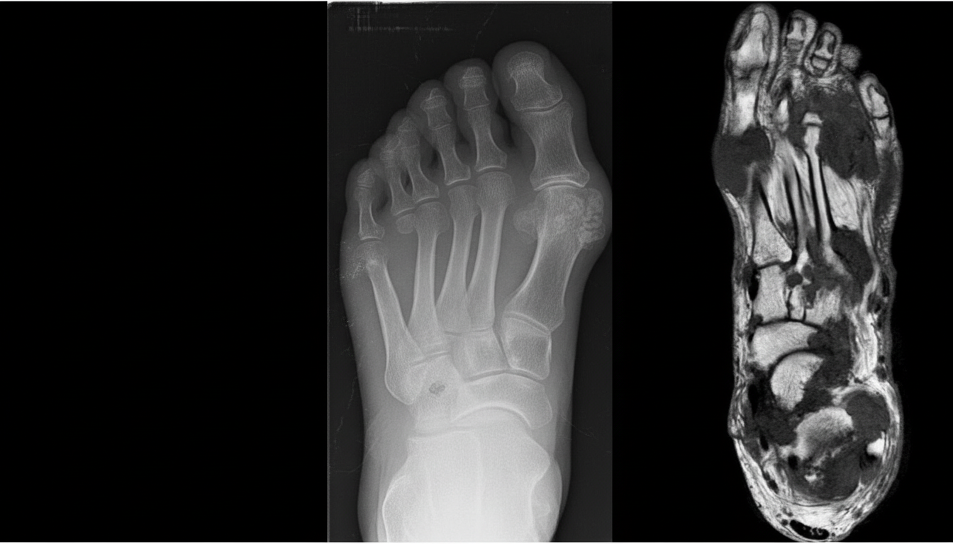
A person presented with swelling of the right 3rd toe. X-ray shows deposition of multiple crystals. A defect in which of the following pathways caused the problem?
Triple bonds are found between which base pairs?
A 3-year-old male child presents with severe self-mutilation behavior (repetitive biting of his lips and fingers causing scarring), choreoathetosis, spasticity, and intellectual disability. Laboratory investigations reveal elevated serum uric acid levels. Which of the following enzymes is likely to be deficient in this child?
Deficiency of purine nucleoside phosphorylase causes which of the following?
The melting temperature of DNA is directly proportional to which of the following?
Nucleic acids absorb UV light maximally at which wavelength?
Which arm of tRNA binds it to the ribosomal surface?
A female patient complains of swelling and pain in her great toe. Her uric acid level is found to be high. A drug that reduces uric acid levels is prescribed. Which of the following enzymes should the drug inhibit?
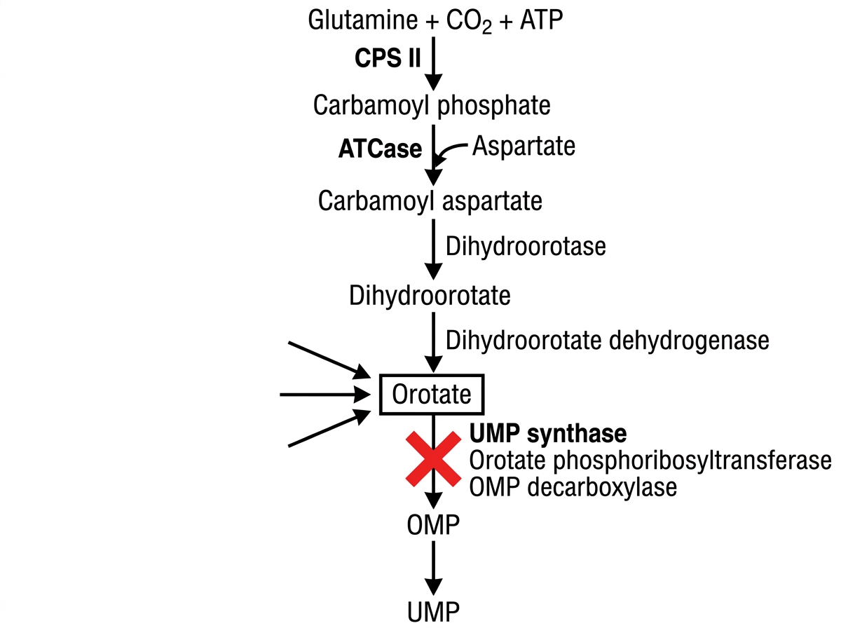
What is the treatment of the condition shown due to absence of enzyme marked as $X$ ?
Which type of bonds are represented by the dotted lines in the image? (AIIMS Nov 2017)

Practice by Chapter
Nucleotide Structure and Function
Practice Questions
DNA Structure and Replication
Practice Questions
RNA Structure and Types
Practice Questions
Transcription: RNA Synthesis
Practice Questions
Post-Transcriptional Modifications
Practice Questions
Translation: Protein Synthesis
Practice Questions
Genetic Code and Codon Usage
Practice Questions
Regulation of Gene Expression
Practice Questions
Mutations and DNA Repair
Practice Questions
Purine Metabolism and Disorders
Practice Questions
Pyrimidine Metabolism and Disorders
Practice Questions
Nucleotide Degradation and Salvage Pathways
Practice Questions
Want unlimited practice?
Get full access to all questions, explanations, and performance tracking.
Scan to download app