
Thalassemias — MCQs

A patient presents with elevated serum iron, low TIBC, and high ferritin. Which of the following conditions is most likely?
A female patient presented with fatigue and a history of piles. Routine complete blood count analysis showed hemoglobin of 9 g/dL, MCV 60fL, and RBC count of 5.2 million. A peripheral smear is provided. Which of the following is the next best investigation after the smear for this patient?

Diagnosis of beta thalassemia is established by what?
Which malformation is associated with mutations in the HOX gene?
Which condition is associated with defects in pre-mRNA splicing and SMN protein dysfunction?
A patient has MCV <80, MCH <23. Which type of anaemia shall be classified?
A 34-year-old, G1P0, presents for genetic counseling at 12 weeks' gestation. The patient has two sisters and a brother; her father has hemophilia. Her siblings are not affected, but she has a nephew who is affected. What is the inheritance pattern of this disorder?
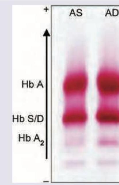
The shown pattern in electrophoresis is due to:
A normal female, whose father is color blind, marries a normal man. What are the chances of their son being color blind?
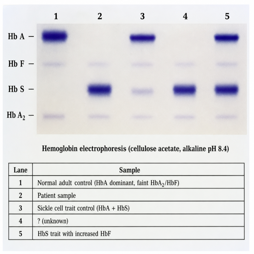
A 19-year-old male of West African descent presents with recurrent episodes of bone pain, acute chest syndrome, and a haemoglobin of 8.2 g/dL with target cells and sickle forms on peripheral smear. His parents are both clinically well. Haemoglobin electrophoresis (cellulose acetate, alkaline pH 8.4) is performed and shown in Image 1. The five lanes are: Lane 1 = Normal adult control (HbA dominant, faint HbA2/HbF); Lane 2 = Patient sample; Lane 3 = Sickle cell trait control (HbA + HbS); Lane 4 = HbC disease control (HbC only); Lane 5 = HbSC disease control (HbS + HbC, roughly equal intensity). Reference positions for HbA, HbS, HbC, and HbA2 are labeled on the gel. Lane 2 shows two bands — one co-migrating with HbS and one co-migrating with HbC — with complete absence of HbA. Based on the electrophoresis pattern in Lane 2, which combination of amino acid substitutions in the beta-globin chain is responsible for the two abnormal haemoglobin variants seen in this patient?
Want unlimited practice?
Get full access to all questions, explanations, and performance tracking.
Scan to download app