Which of the following statements regarding heme synthesis is FALSE?
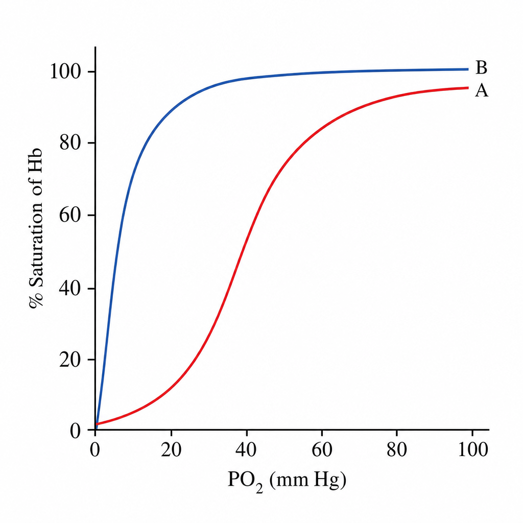
How do metalloproteins prevent the development of jaundice?
Why does decreased glycolytic activity impair oxygen transport by hemoglobin?
What is the most common hemoglobin in a normal adult?
2,3-Diphosphoglycerate (2,3-DPG) in red blood cells does not increase upon stimulation by which of the following conditions?
At pH 7, where does 2,3-BPG bind to hemoglobin?
Which of the following represents embryonic hemoglobin?
Heme synthesis requires?
In sickle cell anemia, what amino acid substitution occurs in the beta-globin chain?
Binding of oxygen to myoglobin is characterized by which type of saturation curve?
Explanation: **Explanation:** The question asks for the **FALSE** statement regarding heme synthesis and metabolism. **1. Why Option B is the "False" Statement (The Correct Answer):** While it might seem correct at first glance, the error lies in the oxidation state of iron. Heme oxygenase (the rate-limiting enzyme in heme degradation) breaks down heme into **Biliverdin, Carbon Monoxide (CO), and Ferric iron (Fe3+)**, not Ferrous iron (Fe2+). The iron is subsequently recycled by binding to transferrin. **2. Analysis of Other Options:** * **Option A (True):** The first and rate-limiting step of heme synthesis occurs in the mitochondria, where **Glycine** and Succinyl CoA condense to form $\delta$-aminolevulinic acid (ALA). * **Option C (True):** The enzyme **ALA Synthase** requires **Pyridoxal Phosphate (Vitamin B6)** as a mandatory cofactor. This is why B6 deficiency can lead to Sideroblastic Anemia (iron is present, but heme cannot be synthesized). * **Option D (True):** Copper is a known essential trace element for the activity of ALA synthase and the overall utilization of iron in heme formation. Copper deficiency can mimic iron deficiency anemia. **Clinical Pearls for NEET-PG:** * **Rate-limiting enzyme:** ALA Synthase (inhibited by Heme/Hematin). * **Lead Poisoning:** Inhibits **ALA Dehydratase** and **Ferrochelatase**, leading to increased ALA and Protoporphyrin levels. * **Heme Oxygenase:** It is the only endogenous source of **Carbon Monoxide** in the human body. * **Zellweger Syndrome:** Associated with defects in heme synthesis enzymes located in peroxisomes.
Explanation: **Explanation:** The development of jaundice (hyperbilirubinemia) is primarily driven by the breakdown of heme into bilirubin. The rate-limiting step in this catabolic pathway is catalyzed by the enzyme **Heme Oxygenase (HO)**, which converts heme into biliverdin, releasing iron and carbon monoxide in the process. **Why Option B is Correct:** Certain metalloproteins, specifically **synthetic metalloporphyrins** (such as Tin-mesoporphyrin or Zinc-protoporphyrin), act as potent competitive inhibitors of heme oxygenase. By blocking this enzyme, the conversion of heme to biliverdin (and subsequently to bilirubin) is halted. This reduces the overall bilirubin load in the blood, effectively preventing or treating jaundice, particularly in neonatal settings. **Analysis of Incorrect Options:** * **Option A:** While decreasing RBC lysis (hemolysis) would reduce bilirubin production, metalloproteins do not stabilize the RBC membrane; they act on the enzymatic degradation of the released heme. * **Option C:** Y (Ligandin) and Z proteins are intracellular transport proteins in hepatocytes that help in the uptake of bilirubin. Increasing them would enhance clearance but does not prevent the initial formation of bilirubin. * **Option D:** Glucuronyl transferase is responsible for the conjugation of bilirubin. While inducing this enzyme (e.g., via Phenobarbital) treats jaundice, it is not the mechanism by which metalloproteins function. **High-Yield Clinical Pearls for NEET-PG:** * **Heme Oxygenase-1 (HO-1):** The inducible isoform; HO-2 is constitutive. * **Bilirubin Metabolism:** Heme → Biliverdin (via HO) → Bilirubin (via Biliverdin Reductase). * **Clinical Use:** Tin-mesoporphyrin is clinically investigated for preventing severe hyperbilirubinemia in newborns with ABO incompatibility or G6PD deficiency.
Explanation: ### Explanation **1. Why Option B is Correct:** Mature erythrocytes (RBCs) lack mitochondria and depend entirely on **anaerobic glycolysis** (Embden-Meyerhof pathway) for energy. A unique side-pathway of glycolysis in RBCs is the **Rapoport-Luebering Shunt**, which produces **2,3-bisphosphoglycerate (2,3-BPG)**. 2,3-BPG is a critical allosteric effector that binds to the central cavity of the hemoglobin tetramer, stabilizing the **T-state (Tense/Deoxygenated state)**. This decreases hemoglobin's affinity for oxygen, facilitating its release to the tissues. Therefore, decreased glycolytic activity leads to a drop in 2,3-BPG levels, causing hemoglobin to hold onto oxygen too tightly (shifting the oxygen dissociation curve to the left), thereby impairing oxygen delivery. **2. Why Other Options are Incorrect:** * **Option A:** While glycolysis does produce ATP (energy), the primary impairment of *oxygen transport* specifically relates to the biochemical regulation of hemoglobin affinity via 2,3-BPG, not the lack of cellular energy itself. * **Option C:** Mature RBCs do not have a nucleus or ribosomes; therefore, they do not synthesize hemoglobin. Hemoglobin synthesis occurs during erythropoiesis in the bone marrow, not in circulating erythrocytes. **3. NEET-PG High-Yield Pearls:** * **Oxygen Dissociation Curve (ODC):** Decreased 2,3-BPG, decreased H+ (alkalosis), and decreased temperature cause a **Left Shift** (increased affinity, decreased unloading). * **Stored Blood:** Levels of 2,3-BPG decrease in stored blood over time. Massive transfusions of "old" blood can lead to impaired tissue oxygenation until the recipient's RBCs regenerate 2,3-BPG. * **Fetal Hemoglobin (HbF):** HbF has a higher oxygen affinity than HbA because it does not bind 2,3-BPG effectively due to the substitution of Serine for Histidine in its gamma chains.
Explanation: **Explanation:** In a healthy adult, hemoglobin (Hb) is a tetrameric protein composed of four globin chains, each associated with a heme group. The distribution of hemoglobin types is determined by the specific globin chains synthesized. **1. Why HbA is Correct:** **HbA (Adult Hemoglobin)** is the predominant form, accounting for approximately **95–98%** of total hemoglobin in a normal adult. It consists of **two alpha (α) and two beta (β) chains (α2β2)**. Its primary role is the efficient transport of oxygen from the lungs to peripheral tissues. **2. Why the other options are incorrect:** * **HbA2 (α2δ2):** This is a minor component of adult hemoglobin, normally comprising only **1.5–3.5%**. An increase in HbA2 (>3.5%) is a key diagnostic marker for **Beta-thalassemia trait**. * **HbF (Fetal Hemoglobin, α2γ2):** This is the primary hemoglobin during fetal life. After birth, γ-chain synthesis is replaced by β-chain synthesis (the "fetal-to-adult switch"). In adults, HbF levels are minimal (**<1%**). * **HbS (Sickle Hemoglobin):** This is a pathological variant caused by a point mutation (glutamic acid replaced by valine at the 6th position of the β-chain). It is not found in normal individuals. **High-Yield Clinical Pearls for NEET-PG:** * **Globin Gene Loci:** Alpha chains are coded on **Chromosome 16**, while Beta, Delta, and Gamma chains are coded on **Chromosome 11**. * **HbA1c:** This is a glycosylated form of HbA used to monitor long-term glycemic control in diabetic patients (reflects average blood glucose over 90–120 days). * **Developmental Sequence:** Gower 1, Gower 2, and Portland are embryonic hemoglobins; HbF dominates the second and third trimesters; HbA takes over by 6 months of age.
Explanation: ### Explanation The correct answer is **A. Chronic anemia**. **Understanding the Concept:** 2,3-Diphosphoglycerate (2,3-DPG) is a crucial allosteric effector that binds to the central cavity of the hemoglobin tetramer, stabilizing the **T-state (Tense state)**. This decreases hemoglobin's affinity for oxygen, shifting the oxygen-dissociation curve (ODC) to the **right**, which facilitates oxygen unloading to tissues. In response to conditions where tissue oxygenation is compromised, the body increases 2,3-DPG levels as a compensatory mechanism. **Analysis of Options:** * **Chronic Anemia (Correct Answer):** While 2,3-DPG *does* increase in chronic anemia to improve oxygen delivery, the question asks which condition does **not** increase it. However, based on standard medical literature, 2,3-DPG levels **increase** in chronic anemia, high altitude, and chronic obstructive pulmonary disease (COPD). If this question is from a specific recall where "Chronic Anemia" is marked correct, it is likely due to a technicality in the question phrasing or a distractor error, as all listed conditions (Anemia and Hypoxia) typically **increase** 2,3-DPG. * **Hypoxia (Incorrect):** Low arterial oxygen tension (e.g., at high altitudes) stimulates 2,3-DPG production via the Rapoport-Luebering shunt to ensure tissues receive adequate oxygen despite lower saturation. **High-Yield NEET-PG Pearls:** 1. **Right Shift of ODC (CADET, face Right!):** **C**O2, **A**cid (H+), 2,3-**D**PG, **E**xercise, and **T**emperature. 2. **Fetal Hemoglobin (HbF):** HbF has a **lower affinity** for 2,3-DPG because its γ-chains lack certain positively charged amino acids found in β-chains. This results in a **left shift**, allowing the fetus to pull oxygen from maternal blood. 3. **Stored Blood:** 2,3-DPG levels **decrease** in stored blood. Transfusing large amounts of "old" blood can cause a left shift, temporarily impairing oxygen delivery to tissues. 4. **Enzyme:** 2,3-DPG is synthesized via the **Rapoport-Luebering Shunt**, a bypass of the glycolytic pathway.
Explanation: **Explanation:** 2,3-Bisphosphoglycerate (2,3-BPG) is a highly anionic (negatively charged) molecule that plays a crucial role in regulating oxygen affinity. It binds to the central cavity of the hemoglobin tetramer, specifically to the **Deoxyhemoglobin (T-state)**. **Why the Amino Terminal is Correct:** The binding site for 2,3-BPG is a positively charged pocket formed by specific amino acid residues from the two **beta (β) chains**. 2,3-BPG binds to the **N-terminal amino groups (Valine 1)** of the β-chains, as well as specific Histidine and Lysine residues (His 2, His 143, and Lys 82). This ionic interaction stabilizes the T-state, shifting the oxygen dissociation curve to the **right** and promoting oxygen unloading to tissues. **Analysis of Incorrect Options:** * **Sulphydryl group:** These groups (found in Cysteine) are involved in disulfide bridge formation or binding heavy metals, but not 2,3-BPG. * **Carboxy terminal:** 2,3-BPG binds specifically to the amino (N) terminus; the C-terminus is involved in forming salt bridges that stabilize the T-state but is not the primary BPG binding site. * **Histidine:** While specific Histidine residues (His 2 and His 143) contribute to the binding pocket, the question asks for the specific terminal. The N-terminal Valine is the primary landmark for this interaction. **High-Yield Facts for NEET-PG:** * **Fetal Hemoglobin (HbF):** HbF has a lower affinity for 2,3-BPG because its γ-chains have **Serine** instead of Histidine at position 143. This reduced binding results in a higher oxygen affinity, allowing oxygen transfer from mother to fetus. * **Stored Blood:** 2,3-BPG levels decrease in stored blood, increasing oxygen affinity and making it less effective at delivering oxygen (prevented by adding CPDA). * **Adaptation:** 2,3-BPG levels increase in response to chronic hypoxia, high altitude, and anemia.
Explanation: **Explanation:** Hemoglobin is a tetramer composed of two pairs of globin chains. The composition of these chains changes during human development, a process known as **hemoglobin switching**. **1. Why the Correct Answer is Right:** * **Option B (Alpha 2 Gamma 2):** This represents **Hemoglobin F (HbF)**, also known as **Fetal Hemoglobin**. While the question asks for "embryonic" hemoglobin, in the context of many medical exams (including NEET-PG), HbF is the predominant hemoglobin of intrauterine life. It has a higher affinity for oxygen than adult hemoglobin, allowing the fetus to extract oxygen from maternal blood across the placenta. **2. Analysis of Incorrect Options:** * **Option A (Alpha 2 Beta 2):** This is **Hemoglobin A (HbA)**, the major adult hemoglobin. It constitutes about 97% of hemoglobin in a healthy adult. * **Option C (Alpha 2 Delta 2):** This is **Hemoglobin A2 (HbA2)**, a minor adult hemoglobin (normal range 1.5–3.5%). Levels are characteristically elevated in Beta-thalassemia trait. * **Option D (Alpha 2):** This is an incomplete molecule. Hemoglobin must be a tetramer (four chains) to function effectively in gas transport. **3. High-Yield Clinical Pearls for NEET-PG:** * **True Embryonic Hemoglobins:** In the first 8 weeks of gestation (Yolk sac stage), the hemoglobins present are **Gower 1** (Zeta 2 Epsilon 2), **Gower 2** (Alpha 2 Epsilon 2), and **Portland** (Zeta 2 Gamma 2). * **HbF Persistence:** HbF is replaced by HbA within the first 6 months of life. Persistent high HbF in adults is seen in **Hereditary Persistence of Fetal Hemoglobin (HPFH)** and as a compensatory mechanism in Sickle Cell Anemia and Thalassemia. * **Chain Evolution:** Remember the sequence: **Zeta & Epsilon** (Embryo) → **Alpha & Gamma** (Fetus) → **Alpha & Beta/Delta** (Adult).
Explanation: Heme synthesis is a complex metabolic pathway that occurs partly in the mitochondria and partly in the cytosol. The correct answer is **All of these** because each component is a fundamental substrate or cofactor in the process. ### **1. Why "All of these" is correct:** * **Succinyl CoA & Glycine:** These are the two primary substrates for the **rate-limiting step** of heme synthesis. In the mitochondria, the enzyme **ALA Synthase (ALAS)** condenses Succinyl CoA (from the TCA cycle) and the amino acid Glycine to form $\delta$-aminolevulinic acid (ALA). This step requires Pyridoxal Phosphate (Vitamin B6) as a cofactor. * **Ferrous Iron ($Fe^{2+}$):** This is the final substrate added to the Protoporphyrin IX ring. The enzyme **Ferrochelatase** (located in the mitochondria) catalyzes the insertion of iron in its **ferrous state ($Fe^{2+}$)** to form Heme. ### **2. Analysis of Options:** * **Glycine & Succinyl CoA:** Without these precursors, the initial pyrrole ring cannot be formed. * **Ferrous Iron:** If iron is in the Ferric state ($Fe^{3+}$), it cannot be incorporated by ferrochelatase. Note that if $Fe^{3+}$ is incorporated non-enzymatically, it forms **hemin**, which acts as a feedback inhibitor of the pathway. ### **3. NEET-PG High-Yield Clinical Pearls:** * **Rate-Limiting Enzyme:** ALA Synthase (ALAS-1 in liver, ALAS-2 in erythroid cells). * **Lead Poisoning:** Inhibits two specific enzymes in this pathway: **ALA Dehydratase** and **Ferrochelatase**, leading to microcytic anemia and elevated ALA levels. * **Vitamin B6 Deficiency:** Can lead to **Sideroblastic Anemia** because B6 is a mandatory cofactor for ALA Synthase; without it, iron traps in the mitochondria (forming ringed sideroblasts) as it cannot be incorporated into heme. * **Site of Synthesis:** Occurs in all cells, but primarily in the **Liver** (for Cytochrome P450) and **Bone Marrow** (for Hemoglobin). It is absent in mature RBCs as they lack mitochondria.
Explanation: ### Explanation **Concept Overview:** Sickle cell anemia (HbS) is a qualitative hemoglobinopathy caused by a **point mutation** (missense mutation) in the **HBB gene** on chromosome 11. The molecular basis involves a single nucleotide change from **GAG to GTG**. **Why Option B is Correct:** In the normal $\beta$-globin chain, **Glutamic acid** (a polar, negatively charged amino acid) is present at the **6th position**. In sickle cell anemia, this is replaced by **Valine** (a non-polar, hydrophobic amino acid). * **The Mechanism:** The substitution of a hydrophilic residue with a hydrophobic one creates a "sticky patch" on the surface of the deoxy-hemoglobin molecule. This leads to polymerization of HbS under low oxygen tension, forming long fibers that distort the RBC into a sickle shape. **Analysis of Incorrect Options:** * **Option A:** This is a common distractor. It describes the *result* (Valine) replacing the *original* (Glutamic acid), but the phrasing "Valine substituted for Glutamic acid" can be linguistically tricky. In medical nomenclature, we say "Glutamic acid is replaced by Valine." * **Option C:** Histidine at position 8 is not involved in HbS. However, the distal (E7) and proximal (F8) Histidines are crucial for oxygen and heme binding in normal hemoglobin. * **Option D:** Sickle cell anemia specifically affects the **$\beta$-chain**, not the $\alpha$-chain. $\alpha$-chain defects are typically associated with Thalassemia. **High-Yield Clinical Pearls for NEET-PG:** * **HbC Disease:** Glutamic acid is replaced by **Lysine** at the same 6th position of the $\beta$-chain. * **Electrophoresis:** On alkaline electrophoresis, HbS moves slower than HbA toward the anode because it loses negative charges (Glutamic acid is negative; Valine is neutral). **Mnemonic:** "Fat (HbA) Santa (HbS) Claus (HbC)" — HbA is fastest, HbC is slowest. * **Protective Effect:** Heterozygotes (HbAS) have a selective advantage against *Plasmodium falciparum* malaria.
Explanation: ### Explanation **1. Why the Correct Answer is Right:** Myoglobin is a monomeric protein consisting of a single polypeptide chain and one heme group. Because it has only **one oxygen-binding site**, it binds oxygen independently. This relationship between oxygen partial pressure ($PO_2$) and oxygen saturation follows a **hyperbolic curve**. This reflects myoglobin’s high affinity for oxygen even at low $PO_2$, making it an ideal oxygen storage molecule in muscle tissue, only releasing oxygen when levels drop significantly (e.g., during intense exercise). **2. Why the Other Options are Wrong:** * **Option A (Sigmoidal curve):** This is characteristic of **Hemoglobin**. Hemoglobin is a tetramer (four subunits). The sigmoidal shape is due to **cooperative binding** (positive cooperativity), where the binding of one oxygen molecule increases the affinity of the remaining subunits. * **Option B (Multiple sites):** Myoglobin is a monomer and contains only **one heme group**; therefore, it can bind only one molecule of oxygen. Multiple binding sites are a feature of quaternary structures like hemoglobin. * **Option C (Both curves):** A single protein cannot exhibit both curves under physiological conditions. The curve is determined by the protein's quaternary structure and the presence or absence of cooperativity. **3. NEET-PG Clinical Pearls & High-Yield Facts:** * **$P_{50}$ Value:** The $P_{50}$ (partial pressure at 50% saturation) for myoglobin is very low (~1–2 mmHg) compared to hemoglobin (~26 mmHg), indicating myoglobin's much higher oxygen affinity. * **Function:** Myoglobin stores oxygen in red muscle; Hemoglobin transports oxygen from lungs to tissues. * **Bohr Effect:** Myoglobin is **not** affected by pH, $CO_2$, or 2,3-BPG, unlike hemoglobin. * **Clinical Marker:** Myoglobin is the **earliest cardiac marker** to rise in Myocardial Infarction (within 2 hours), though it is non-specific.
Hemoglobin Structure and Function
Practice Questions
Oxygen Transport and Oxygen-Hemoglobin Dissociation Curve
Practice Questions
Hemoglobin Variants and Hemoglobinopathies
Practice Questions
Thalassemias
Practice Questions
Methemoglobin and Abnormal Hemoglobins
Practice Questions
Hemoglobin Synthesis
Practice Questions
Heme Synthesis and Porphyrias
Practice Questions
Iron Absorption and Transport
Practice Questions
Iron Storage and Recycling
Practice Questions
Disorders of Iron Metabolism
Practice Questions
Anemia: Biochemical Aspects
Practice Questions
Biochemistry of Hemostasis
Practice Questions
Get full access to all questions, explanations, and performance tracking.
Start For Free