Trinucleotide sequence associated with spinocerebellar ataxia is?
What is the most definitive method for confirming 46,XY disorders of sexual development?
Pendred syndrome is caused by a mutation in which gene?
Gene responsible for Wilson disease is situated on which chromosome?
Which type of genetic disorders are more likely to affect boys?
Which of the following statements is true regarding hemophilia inheritance?
In argininosuccinase deficiency, what should be supplemented to continue the urea cycle ?
An infant is brought by his parents with complaints that his urine turns black on standing. Which of the following metabolic disorders is likely?
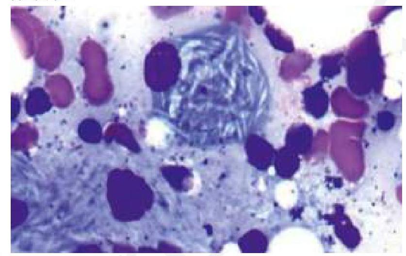
A child presents with bone pain and hepatosplenomegaly, indicative of Gaucher's disease. A trephine biopsy and aspirate show the following finding. Which of the following is the most likely enzyme deficient in this condition?

A 4-year-old boy from a first-degree consanguineous couple presents with darkening of the urine to an almost black color when left standing. He has a normal sibling and no other medical problems. His growth and development are normal. Which of the following metabolites is most likely to be elevated in this patient?
Explanation: ***CAG*** - The **CAG trinucleotide repeat** is associated with **most forms of spinocerebellar ataxia (SCA)**, including SCA1, SCA2, SCA3 (Machado-Joseph disease), SCA6, SCA7, and SCA17. - An expansion of this repeat leads to an elongated **polyglutamine tract** in the affected protein, causing protein misfolding and neuronal dysfunction. - CAG is the **most common** trinucleotide repeat expansion associated with SCA. *CUG* - **CUG repeats** are primarily associated with **myotonic dystrophy type 1**. - While CUG expansions are found in **SCA8**, CAG repeats are far more commonly associated with the majority of spinocerebellar ataxias. - The expansion of CUG repeats in the **3' untranslated region** causes RNA-mediated toxicity. *GGC* - **GGC repeats** are not a common trinucleotide expansion associated with classic spinocerebellar ataxia. - While other repeat expansions exist in various genetic disorders, GGC is not the primary repeat linked to SCA. *CGG* - **CGG trinucleotide repeat** expansions are characteristic of **Fragile X syndrome**, a common cause of inherited intellectual disability. - This repeat expansion occurs in the FMR1 gene and is not the primary repeat associated with spinocerebellar ataxia.
Explanation: ***Genetic testing*** - Genetic testing, such as **karyotyping** and **DNA sequencing**, can identify abnormalities in genes responsible for male sexual development, confirming 46,XY DSD. - This method directly pinpoints the underlying genetic cause, providing a **definitive diagnosis** for specific DSDs like **androgen insensitivity syndrome** or **5-alpha-reductase deficiency**. - Molecular analysis can detect mutations in genes like **SRY, AR, SRD5A2**, and other genes involved in sex determination and differentiation. *USG abdomen* - **Ultrasonography** of the abdomen can visualize internal reproductive organs and identify anatomical abnormalities, but it does not reveal the underlying genetic cause. - While useful for assessing morphology, it is an **imaging modality** and not a definitive diagnostic tool for genetic conditions. *Hormonal study* - **Hormonal studies** measure levels of androgens, estrogens, and other related hormones, which can indicate impaired hormone production or action. - While essential for understanding the **physiological impact** of DSDs, hormonal studies provide indirect evidence and do not definitively identify the genetic origin. *Gonadal biopsy* - **Histopathological examination** of gonadal tissue can reveal tissue architecture and cell types, which may support the diagnosis. - However, it is an **invasive procedure** and does not identify the specific genetic mutation responsible for the DSD, making it less definitive than genetic testing.
Explanation: ***SLC26A4 gene*** - Pendred syndrome is an autosomal recessive disorder characterized by **sensorineural hearing loss** and **goiter**, caused by mutations in the *SLC26A4 gene*. - The *SLC26A4 gene* encodes pendrin, a **chloride-iodide transporter** found in the inner ear and thyroid gland, crucial for endolymph production and iodide transport. *Bartillin* - The *BSND gene* encodes bartillin, which is associated with **Bartter syndrome type IV**, characterized by renal salt wasting, deafness, and metabolic alkalosis. - While it involves deafness, it's distinct from Pendred syndrome as it doesn't primarily involve thyroid dysfunction. *Fibrillin* - **Fibrillin-1** is encoded by the *FBN1 gene* and mutations in this gene cause **Marfan syndrome**, a genetic disorder affecting connective tissue, leading to cardiovascular, skeletal, and ocular abnormalities. - It is not associated with hearing loss or thyroid issues, as seen in Pendred syndrome. *Reticulin* - Reticulin refers to a type of collagen fiber (primarily **collagen type III**) found in connective tissues. - Mutations directly related to the protein "reticulin" are not known to cause specific genetic syndromes like Pendred syndrome.
Explanation: ***Chromosome 13*** - The **ATP7B gene**, responsible for Wilson disease, is located on **chromosome 13** (specifically 13q14.3). - Mutations in this gene lead to impaired copper transport and accumulation in various organs, particularly the liver and brain. *Chromosome 11* - This chromosome is associated with various genetic disorders, such as **sickle cell disease** (HBB gene) and **Beckwith-Wiedemann syndrome**. - Wilson disease is not associated with genes on this chromosome. *Chromosome 12* - **Phenylketonuria (PKU)** (PAH gene) and some forms of **Noonan syndrome** are linked to genes on chromosome 12. - Wilson disease is not associated with genes on this chromosome. *Chromosome 14* - This chromosome contains genes associated with **Alpha-1 antitrypsin deficiency** and some forms of **Alzheimer's disease**. - It does not contain the ATP7B gene responsible for Wilson disease.
Explanation: ***X-linked Recessive*** - X-linked recessive disorders are more likely to affect boys because they have only one X chromosome. If that X chromosome carries the affected gene, they will express the disorder. - Females have two X chromosomes, so if one carries the affected gene, the other typically healthy X chromosome can compensate, making them carriers but often asymptomatic. *Autosomal Dominant* - These disorders affect individuals when they inherit just one copy of a mutated gene on an autosome (non-sex chromosome). - They affect both males and females equally since autosomes are not sex-linked. *Autosomal Recessive* - These disorders require two copies of a mutated gene on an autosome for the disease to manifest. - They affect both males and females equally because the genes are on non-sex chromosomes. *X-linked Dominant* - These disorders are caused by a mutation on an X chromosome, where only one copy of the mutated gene is sufficient to cause the disorder. - While they can affect both males and females, females are usually more commonly affected and often present with milder symptoms, while affected males typically have more severe phenotypes.
Explanation: ***Females can be carriers, and males are typically affected.*** - Hemophilia is an **X-linked recessive disorder**, meaning the gene responsible is located on the X chromosome. - Males have only one X chromosome, so if they inherit the affected gene, they will express the disorder, while females have two X chromosomes, allowing them to be **asymptomatic carriers** if only one X chromosome carries the mutation. - This option accurately describes the **typical inheritance pattern** where carrier females (heterozygous) transmit the condition to their sons who become affected. *Females can be carriers, and some males may be unaffected.* - While this statement is technically true, it is less precise for describing hemophilia inheritance patterns. - The focus on "some males may be unaffected" is less informative than emphasizing that males who inherit the mutation are **typically affected**, which is the key clinical characteristic. - This option doesn't emphasize the primary pattern that makes X-linked recessive inheritance clinically significant. *Males are carriers, and females are typically affected.* - Males cannot be carriers for X-linked recessive disorders; if a male carries the gene on his single X chromosome, **he will be affected**, not a carrier. - **Females are rarely affected** as they would need to inherit the mutated gene from both parents (an affected father and a carrier/affected mother), which is uncommon. - This statement reverses the actual inheritance pattern. *All males are affected, and females cannot be carriers.* - It is incorrect that all males are affected; only males who **inherit the specific defective X chromosome** will be affected. - Females **can and often are carriers**, as they possess two X chromosomes, and one can carry the mutated gene while the other compensates through X-inactivation (lyonization).
Explanation: ***Correct: Arginine*** - **Arginine** supplementation is the primary treatment for argininosuccinase deficiency - Provides the product of the blocked reaction, allowing the urea cycle to continue - Maintains essential arginine levels and enables **nitrogen scavenger pathway** - Accumulated **argininosuccinate** is excreted in urine, removing excess nitrogen - This bypasses the deficient enzyme while facilitating alternative nitrogen disposal *Incorrect: Aspartate* - **Aspartate** combines with citrulline to form argininosuccinate via argininosuccinate synthetase - Supplementing aspartate alone would increase argininosuccinate accumulation without resolving the enzymatic block - Does not provide the end product (arginine) needed to continue the urea cycle - Not part of the primary therapeutic strategy for this deficiency *Incorrect: Citrulline* - **Citrulline** is actually used as adjunct therapy alongside arginine in argininosuccinase deficiency - However, citrulline supplementation alone cannot **continue the urea cycle** because the enzyme block prevents conversion of argininosuccinate to arginine - While it helps generate argininosuccinate for urinary excretion (nitrogen scavenging), it doesn't provide arginine needed for urea formation - **Arginine** remains the primary supplement as it directly supplies the missing product and enables cycle completion *Incorrect: Argininosuccinate* - **Argininosuccinate** accumulates in this deficiency due to the enzyme block - Supplementing the substrate of a deficient enzyme would worsen metabolic accumulation - The therapeutic goal is to facilitate its excretion, not increase its levels - Contraindicated in this condition
Explanation: ***Alkaptonuria*** - **Alkaptonuria** is an autosomal recessive disorder characterized by a deficiency of **homogentisate 1,2-dioxygenase**, an enzyme involved in the metabolism of tyrosine. - The accumulation of **homogentisic acid** in tissues and urine causes the urine to turn black on standing due to oxidation. *Phenylketonuria* - **Phenylketonuria (PKU)** is caused by a deficiency of **phenylalanine hydroxylase**, leading to the accumulation of phenylalanine. - While it can manifest with intellectual disability and neurological symptoms, it does not typically cause the urine to turn black. *Homocystinuria* - **Homocystinuria** is a disorder of methionine metabolism, typically due to a deficiency of **cystathionine beta-synthase**. - It is characterized by intellectual disability, skeletal abnormalities, and lens dislocation, but not black urine. *Maple syrup urine disease* - **Maple syrup urine disease (MSUD)** results from a deficiency of **branched-chain alpha-keto acid dehydrogenase complex**, leading to the accumulation of branched-chain amino acids. - The distinguishing feature is urine that smells like maple syrup, not turning black.
Explanation: ***Correct: Glucocerebrosidase*** - The clinical presentation of **bone pain**, **hepatosplenomegaly**, and the characteristic histological finding of **lipid-laden macrophages** (Gaucher cells) with a **crinkled paper** appearance in the bone marrow aspirate are highly suggestive of **Gaucher's disease**. - **Gaucher's disease** is caused by a deficiency of the lysosomal enzyme **glucocerebrosidase**, leading to the accumulation of **glucocerebroside**. *Incorrect: Hexosaminidase* - Deficiency of **hexosaminidase A** is associated with **Tay-Sachs disease**, which presents with neurological degeneration but typically **lacks hepatosplenomegaly** and bone pain. - The histological findings in Tay-Sachs disease would show neuronal storage of **GM2 gangliosides**, not Gaucher cells. *Incorrect: Sphingomyelinase* - Deficiency of **sphingomyelinase** causes **Niemann-Pick disease**, characterized by hepatosplenomegaly, neurological involvement, and interstitial lung disease, but the storage cells (foam cells) have a **foamy appearance** due to sphingomyelin accumulation, not the "crinkled paper" appearance of Gaucher cells. - While there is organomegaly, the distinct **histological features** in the image rule out Niemann-Pick disease. *Incorrect: Alpha 1,4-glucosidase* - Deficiency of **alpha 1,4-glucosidase** (acid maltase) causes **Pompe disease** (Glycogen Storage Disease Type II), which primarily affects muscle and liver with **glycogen accumulation**. - Pompe disease does not typically present with the same type of **bone pain** or the characteristic **Gaucher cells** seen in the image.
Explanation: ***Correct: Homogentisate*** - The clinical presentation of urine darkening to an almost black color upon standing, especially in a child from a consanguineous union, is classic for **alkaptonuria**. - **Alkaptonuria** is an autosomal recessive disorder caused by a deficiency of homogentisate 1,2-dioxygenase, leading to the accumulation of **homogentisate** in the body. - The homogentisic acid polymerizes upon exposure to air, causing the characteristic dark discoloration of urine. - Patients are typically asymptomatic in childhood; **ochronosis** (pigment deposition in connective tissues) and arthritis develop in adulthood. *Incorrect: Methylmalonate* - Elevated **methylmalonate** is characteristic of **methylmalonic aciduria**, a disorder of branched-chain amino acid metabolism or vitamin B12 deficiency. - This condition typically presents with metabolic acidosis, developmental delays, and failure to thrive, which are not described in this patient. *Incorrect: Phenylpyruvate* - Elevated **phenylpyruvate** is a hallmark of **phenylketonuria (PKU)**, an inborn error of metabolism affecting phenylalanine breakdown. - Untreated PKU leads to severe neurological impairment and intellectual disability, which are absent in this child. *Incorrect: α-Ketoisovalerate* - Elevated **α-Ketoisovalerate** is one of the branched-chain keto acids found in **maple syrup urine disease (MSUD)**. - MSUD presents in infancy with poor feeding, lethargy, and a characteristic maple syrup odor in the urine, not darkening urine.
Single Gene Disorders
Practice Questions
Biochemical Diagnosis of Genetic Disorders
Practice Questions
Inborn Errors of Metabolism
Practice Questions
Lysosomal Storage Diseases
Practice Questions
Glycogen Storage Diseases
Practice Questions
Disorders of Lipoprotein Metabolism
Practice Questions
Disorders of Purine and Pyrimidine Metabolism
Practice Questions
Hemoglobinopathies
Practice Questions
Porphyrias
Practice Questions
Biochemical Markers for Disease Diagnosis
Practice Questions
Newborn Screening for Genetic Disorders
Practice Questions
Enzyme Replacement Therapy
Practice Questions
Get full access to all questions, explanations, and performance tracking.
Start For Free