A severely retarded infant presents with hepatosplenomegaly and a cherry-red spot in the macula. 
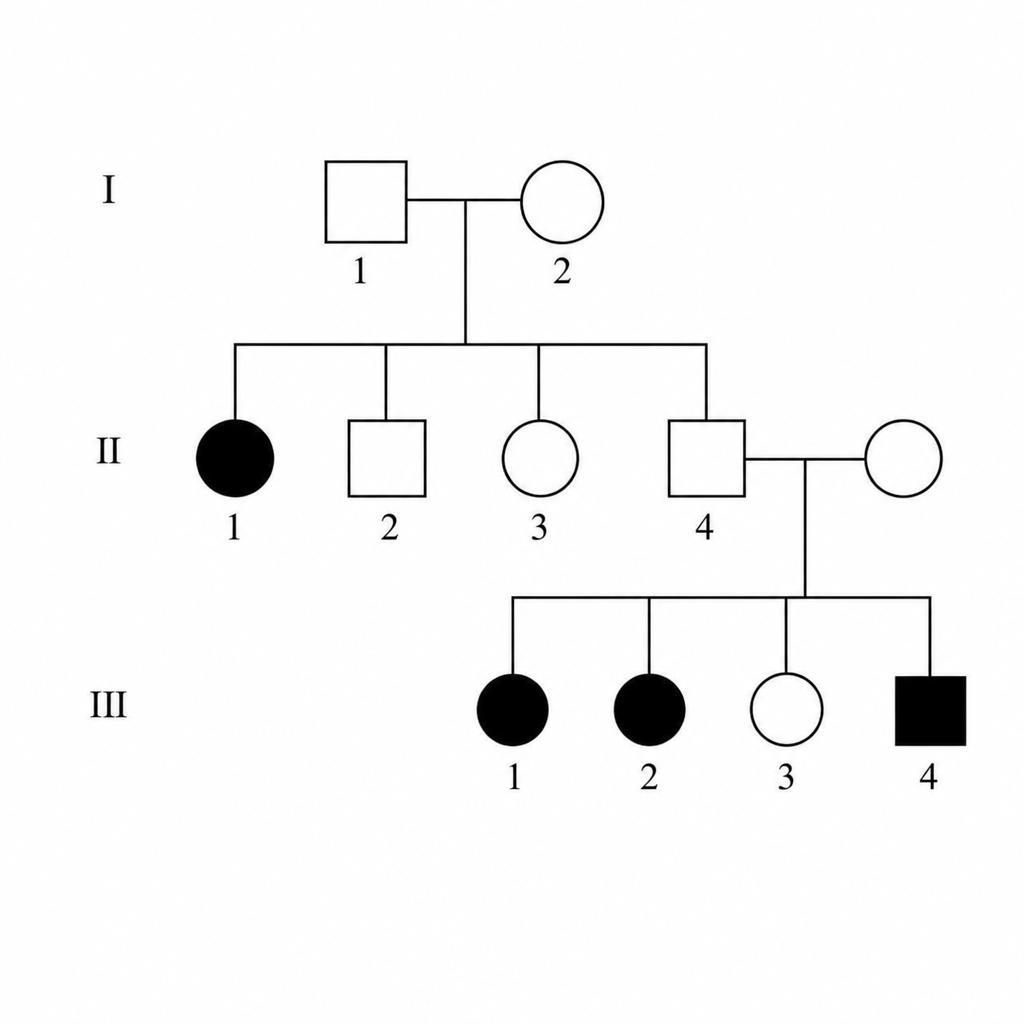
Which of the following is the most likely cause of these findings?
Gene therapy is successful for the treatment of which of the following conditions?
Deficiency of which of the following enzymes leads to toxicity of 5-fluorouracil?
What is characteristically seen in Hartnup disorder?
All of the following are characteristic of I-cell disease except?
Which of the following are disorders of copper metabolism?
A 19-year-old male presents for a routine physical examination for sports activities. His fasting blood glucose is 7.5 mM. C-peptide and insulin levels are near normal under fasting conditions. Postprandially, his blood glucose levels are only slightly elevated before stabilizing at fasting levels. He denies excessive thirst or urination but recalls his mother had gestational diabetes. What type of inheritance best characterizes this alteration in glucose homeostasis?
What is the most serious complication of acute hemolytic anemia in G6PD deficiency?
Gaucher's disease is due to a deficiency of which enzyme?
In which of the following conditions is the formation of a calcium-containing kidney stone NOT likely?
Explanation: **Explanation:** The clinical triad of **neurodegeneration (retardation), cherry-red spot, and hepatosplenomegaly** is the classic presentation of **Niemann-Pick Disease (Type A)**. This autosomal recessive lysosomal storage disorder is caused by a deficiency of **sphingomyelinase**, leading to the accumulation of sphingomyelin in the brain, liver, and spleen. **Why the other options are incorrect:** * **Tay-Sachs Disease:** While it presents with neurodegeneration and a **cherry-red spot**, it is characterized by the **absence of hepatosplenomegaly**. This is the most critical clinical differentiator from Niemann-Pick. It is caused by Hexosaminidase A deficiency. * **Gaucher’s Disease:** This is the most common lysosomal storage disease and features massive hepatosplenomegaly and bone crises (Erlenmeyer flask deformity), but it **does not** typically present with a cherry-red spot. * **Metachromatic Leukodystrophy:** Caused by Arylsulfatase A deficiency, it leads to central and peripheral demyelination (ataxia, dementia). It does not present with a cherry-red spot or significant organomegaly. **NEET-PG High-Yield Pearls:** 1. **The "No-Spleen" Rule:** If you see a cherry-red spot **without** hepatosplenomegaly, think **Tay-Sachs**. If organomegaly is **present**, think **Niemann-Pick**. 2. **Histology:** Niemann-Pick is associated with **"Foam cells"** (lipid-laden macrophages), whereas Gaucher’s features **"Crumpled tissue paper"** appearance (Gaucher cells). 3. **Cherry-red spot differential:** Also seen in Central Retinal Artery Occlusion (CRAO), Sandhoff disease, and Sialidosis.
Explanation: **Explanation:** **Adenosine Deaminase (ADA) Deficiency** is the correct answer because it was the first disease ever treated with gene therapy (1990). ADA deficiency is a form of **Severe Combined Immunodeficiency (SCID)** characterized by the accumulation of deoxyadenosine, which is toxic to T and B lymphocytes. In gene therapy, functional ADA genes are inserted into the patient’s hematopoietic stem cells or T-cells using viral vectors (like retroviruses), restoring immune function. **Analysis of Incorrect Options:** * **Krabbe’s Disease:** This is a lysosomal storage disorder caused by galactocerebrosidase deficiency. While research is ongoing, standard treatment remains hematopoietic stem cell transplantation (HSCT) rather than established gene therapy. * **Hodgkin’s Lymphoma:** This is a malignancy of the lymphoid tissue. It is primarily treated with chemotherapy (e.g., ABVD regimen) and radiotherapy. Gene therapy is not a standard clinical modality for this cancer. * **Beta Thalassemia:** While gene therapy (using Lentiglobin) has recently shown promise and received some regulatory approvals, it is not the "classic" or historically successful example cited in medical examinations compared to ADA-SCID. **NEET-PG High-Yield Pearls:** * **First Gene Therapy:** Performed by William French Anderson in 1990 on Ashanti DeSilva (4-year-old with ADA-SCID). * **Biochemical Hallmark:** Deficiency of ADA leads to increased **dATP levels**, which inhibits **ribonucleotide reductase**, preventing DNA synthesis and causing lymphotoxicity. * **Inheritance:** ADA deficiency is **Autosomal Recessive**, whereas the most common form of SCID (IL-2 receptor gamma chain mutation) is X-linked.
Explanation: **Explanation:** The correct answer is **Dihydropyrimidine dehydrogenase (DPD)**. **Why DPD is the correct answer:** 5-Fluorouracil (5-FU) is a pyrimidine analog used in cancer chemotherapy. Under normal physiological conditions, more than 80% of administered 5-FU is catabolized and inactivated in the liver. **Dihydropyrimidine dehydrogenase (DPD)** is the rate-limiting enzyme in this catabolic pathway, responsible for converting 5-FU into its inactive metabolite, dihydrofluorouracil (DHFU). Patients with **DPD deficiency** cannot efficiently clear the drug, leading to an accumulation of toxic levels of 5-FU. This results in severe, potentially fatal toxicities, including profound myelosuppression, mucositis, and neurotoxicity. **Why the other options are incorrect:** * **A. Purine nucleoside phosphorylase (PNP):** This enzyme is involved in the purine salvage pathway. Its deficiency leads to T-cell immunodeficiency but has no role in 5-FU metabolism. * **C. PRPP synthase:** This enzyme catalyzes the formation of phosphoribosyl pyrophosphate (PRPP), a key precursor for both purine and pyrimidine synthesis. Overactivity leads to gout; it is not involved in 5-FU catabolism. * **D. Adenosine deaminase (ADA):** ADA deficiency leads to Severe Combined Immunodeficiency (SCID) due to the accumulation of dATP, which inhibits ribonucleotide reductase. It is specific to purine metabolism. **High-Yield Clinical Pearls for NEET-PG:** * **Mechanism of Action:** 5-FU acts primarily by inhibiting **Thymidylate Synthase**, leading to "thymineless death" of cells. * **Antidote:** **Uridine triacetate** is the FDA-approved antidote for 5-FU overdose or severe toxicity. * **Pharmacogenomics:** Screening for *DPYD* gene mutations is increasingly recommended before starting 5-FU or Capecitabine to prevent severe adverse reactions.
Explanation: **Explanation:** **Hartnup disorder** is an autosomal recessive metabolic disorder caused by a mutation in the **SLC6A19 gene**, which encodes a sodium-dependent neutral amino acid transporter. This defect leads to the malabsorption of neutral amino acids (most importantly **Tryptophan**) from the small intestine and a failure of their reabsorption in the proximal renal tubules. **Why Option A is correct:** Tryptophan is an essential precursor for the endogenous synthesis of **Niacin (Vitamin B3)**. In Hartnup disease, the profound deficiency of Tryptophan results in secondary Niacin deficiency. This manifests clinically as **Pellagra**, characterized by the "3 Ds": Dermatitis (a photosensitive, scaly rash), Diarrhea, and Dementia. The rash is typically seen on sun-exposed areas (e.g., Casal’s necklace). **Why other options are incorrect:** * **B. Burned feet rash:** This is associated with **Pantothenic acid (Vitamin B5)** deficiency, characterized by paresthesia and burning sensations in the lower extremities. * **C. Alopecia:** While seen in Zinc deficiency or Biotin deficiency, it is not a primary feature of Hartnup disorder. * **D. Vitiligo:** This is an autoimmune destruction of melanocytes and is unrelated to amino acid transport defects. **High-Yield Clinical Pearls for NEET-PG:** * **Diagnosis:** Confirmed by detecting **neutral aminoaciduria** (specifically Alanine, Serine, Threonine, Valine, Leucine, Isoleucine, Phenylalanine, Tyrosine, and Tryptophan) in a 24-hour urine sample. * **Treatment:** High-protein diet and **Nicotinic acid/Nicotinamide** supplementation. * **Differential:** Unlike dietary Pellagra, Hartnup disorder will show high levels of neutral amino acids in the urine.
Explanation: **Explanation:** **I-cell disease (Inclusion Cell disease or Mucolipidosis II)** is a severe lysosomal storage disorder caused by a deficiency of the enzyme **N-acetylglucosamine-1-phosphotransferase**. This enzyme is responsible for phosphorylating mannose residues on nascent lysosomal enzymes in the Golgi apparatus. Without the **Mannose-6-Phosphate (M6P)** tag, these enzymes are not targeted to lysosomes but are instead constitutively secreted into the extracellular space. **Why Buphthalmos is the correct answer:** Buphthalmos (enlargement of the eyeball, typically seen in congenital glaucoma) is **not** a feature of I-cell disease. While I-cell disease involves significant ocular pathology, the classic finding is **corneal clouding**. Buphthalmos is more characteristically associated with conditions like Sturge-Weber syndrome or primary congenital glaucoma. **Analysis of incorrect options:** * **Coarse facial features (A):** This is a hallmark of I-cell disease, similar to Hurler syndrome but often presenting earlier and more severely. It is due to the accumulation of undegraded glycosaminoglycans and lipids in tissues. * **Corneal clouding (B):** This occurs due to the deposition of storage material in the corneal stroma. It is a key clinical differentiator from Hunter syndrome (where it is absent). * **Failure to thrive (C):** Patients typically show severe growth retardation and developmental delays starting in early infancy due to systemic cellular dysfunction. **NEET-PG High-Yield Pearls:** * **Biochemical Hallmark:** Elevated levels of lysosomal enzymes in the **plasma** and decreased levels within the **cells** (lysosomes). * **Microscopy:** Presence of **intracytoplasmic inclusions** (I-cells) in fibroblasts. * **Clinical Comparison:** Often described as "Hurler syndrome on steroids" because it presents with similar features (skeletal deformities, organomegaly) but at birth or in early infancy. * **Inheritance:** Autosomal Recessive.
Explanation: **Explanation:** The correct answer is **D. All of the above**, as all three conditions are caused by mutations in genes responsible for copper transport and homeostasis. 1. **Wilson’s Disease (Hepatolenticular Degeneration):** Caused by a mutation in the **ATP7B** gene on chromosome 13. This leads to impaired biliary excretion of copper and failure to incorporate copper into ceruloplasmin. The result is toxic copper accumulation in the **liver, brain (basal ganglia), and cornea (Kayser-Fleischer rings)**. 2. **Menkes Kinky Hair Syndrome:** Caused by a mutation in the **ATP7A** gene. Unlike Wilson’s, this involves defective intestinal absorption and systemic transport of copper. This leads to **copper deficiency** in tissues, affecting copper-dependent enzymes like lysyl oxidase (causing brittle, "kinky" hair and connective tissue defects) and tyrosinase. 3. **MEDNIK Syndrome:** This is a rare neurocutaneous syndrome caused by mutations in the **AP1S1** gene, which affects the trafficking of both **ATP7A and ATP7B** transporters. It presents with a unique clinical combination of Mental retardation, Enteropathy, Deafness, Neuropathy, Ichthyosis, and Keratoderma (hence the acronym). **High-Yield Clinical Pearls for NEET-PG:** * **ATP7A vs. ATP7B:** Remember **"A"** for **A**bsorption (Menkes) and **"B"** for **B**iliary excretion (Wilson’s). * **Biochemical Markers:** In Wilson’s, look for **decreased** serum ceruloplasmin and **increased** 24-hour urinary copper excretion. * **Enzyme Link:** The "kinky hair" in Menkes is specifically due to defective **Lysyl Oxidase**, which requires copper for cross-linking collagen and elastin. * **Treatment:** Wilson’s is treated with copper chelators (D-Penicillamine, Trientine) and Zinc (which inhibits intestinal absorption).
Explanation: ### Explanation **1. Why Autosomal Dominant is Correct:** The clinical presentation is classic for **MODY (Maturity-Onset Diabetes of the Young)**, specifically **MODY-2**, which is caused by a mutation in the **Glucokinase (GCK) gene**. * **The Mechanism:** Glucokinase acts as the "glucose sensor" in pancreatic beta cells. A mutation increases the threshold for insulin release, leading to mild, stable, non-progressive fasting hyperglycemia (typically 5.5–8.0 mmol/L). * **The Inheritance:** All forms of MODY (including the most common types: HNF1A/MODY-3 and GCK/MODY-2) follow an **Autosomal Dominant** inheritance pattern. The mention of the mother having "gestational diabetes" (often misdiagnosed MODY-2) further supports a vertical transmission pattern. **2. Why Incorrect Options are Wrong:** * **Autosomal Recessive:** Rare forms of neonatal diabetes can be recessive, but MODY presents in adolescence/young adulthood and requires only one affected allele for the phenotype. * **Sex-linked:** There are no common primary disorders of glucose homeostasis linked to the X or Y chromosomes. * **Mitochondrial:** While Maternal Inherited Diabetes and Deafness (MIDD) exists, it is characterized by hearing loss and a different clinical progression, not the stable, asymptomatic hyperglycemia seen here. **3. Clinical Pearls for NEET-PG:** * **MODY-2 (Glucokinase):** Mildest form; usually asymptomatic; requires no treatment; no long-term microvascular complications. * **MODY-3 (HNF1A):** Most common type; progressive; high risk of complications; extremely sensitive to **Sulfonylureas**. * **Key Differentiator:** Unlike Type 1 DM, MODY patients have **detectable C-peptide** (insulin production is preserved) and lack autoantibodies. Unlike Type 2 DM, they are usually young, non-obese, and have no insulin resistance.
Explanation: **Explanation:** **Glucose-6-Phosphate Dehydrogenase (G6PD) deficiency** is an X-linked recessive disorder where the lack of G6PD enzyme leads to decreased NADPH production, making erythrocytes vulnerable to oxidative stress. **Why Acute Renal Failure (ARF) is the correct answer:** During an acute hemolytic crisis (triggered by fava beans, infections, or drugs like Primaquine), there is massive intravascular hemolysis. This releases a large amount of **free hemoglobin** into the plasma. The hemoglobin is filtered by the glomeruli and enters the renal tubules. In the acidic environment of the distal tubules, hemoglobin precipitates and forms casts, leading to **Acute Tubular Necrosis (ATN)**. Additionally, the release of heme causes direct oxidative damage to tubular cells and induces renal vasoconstriction, ultimately resulting in **Acute Renal Failure**. This is the most life-threatening acute complication of the hemolytic episode. **Why other options are incorrect:** * **B. Congestive cardiac failure:** While severe chronic anemia can lead to high-output heart failure, it is rarely the immediate "most serious" complication of an acute G6PD hemolytic episode compared to renal shutdown. * **C. Cerebral infarction:** Hemolysis does not typically cause arterial occlusion or stroke; this is more characteristic of Sickle Cell Disease (due to vaso-occlusive crises). * **D. Acute liver failure:** While hemolysis increases unconjugated bilirubin (leading to jaundice), it does not cause hepatocytes necrosis or functional liver failure. **High-Yield Clinical Pearls for NEET-PG:** * **Hallmark Histology:** **Heinz bodies** (denatured hemoglobin) and **Bite cells** (degluticytes) formed by splenic macrophages. * **Common Triggers:** Infections (most common), Fava beans (Favism), and drugs (Sulfonamides, Primaquine, Nitrofurantoin). * **Diagnostic Note:** G6PD enzyme levels may be **falsely normal** during an acute attack because older cells (with low enzyme levels) have lysed, leaving only younger reticulocytes with higher enzyme activity. Testing should be repeated after 6–8 weeks.
Explanation: **Explanation:** **Gaucher’s disease** is the most common lysosomal storage disorder. It is inherited in an autosomal recessive pattern and is caused by a deficiency of the enzyme **$\beta$-glucosidase** (also known as **glucocerebrosidase**). 1. **Why Option B is Correct:** Under normal physiological conditions, $\beta$-glucosidase cleaves glucocerebroside into glucose and ceramide. A deficiency leads to the accumulation of **glucocerebroside** within the lysosomes of macrophages. These lipid-laden macrophages transform into characteristic **"Gaucher cells,"** which classically appear as having "wrinkled tissue paper" cytoplasm. 2. **Why Other Options are Incorrect:** * **Option A (Sphingomyelinase):** Deficiency leads to **Niemann-Pick disease**, characterized by hepatosplenomegaly and "foam cells." * **Option C (Hexosaminidase-A):** Deficiency leads to **Tay-Sachs disease**, characterized by a cherry-red spot on the macula but *no* hepatosplenomegaly. * **Option D ($\beta$-Galactosidase):** Deficiency leads to **Krabbé disease**, characterized by the presence of globoid cells and severe demyelination. **High-Yield Clinical Pearls for NEET-PG:** * **Clinical Triad:** Hepatosplenomegaly, bone involvement (Erlenmeyer flask deformity of the femur, bone crises), and pancytopenia. * **Biomarker:** Elevated levels of **serum acid phosphatase** (tartrate-resistant) and **chitotriosidase** are often seen. * **Treatment:** Enzyme Replacement Therapy (ERT) with recombinant enzymes like **Imiglucerase** is the gold standard. * **Note:** Unlike many other sphingolipidoses, Gaucher Type 1 (the most common form) is non-neuronopathic.
Explanation: **Explanation:** The formation of kidney stones is primarily determined by the chemical composition of the solute in excess. To answer this question, one must distinguish between **Hypercalcemia** (leading to calcium stones) and **Hyperuricemia** (leading to urate stones). **Why Lesch-Nyhan Syndrome is the correct answer:** Lesch-Nyhan syndrome is an X-linked recessive disorder caused by a deficiency of the enzyme **HGPRT** (Hypoxanthine-Guanine Phosphoribosyltransferase). This deficiency leads to the failure of the purine salvage pathway, resulting in the massive overproduction of **Uric Acid**. Consequently, patients develop **Uric Acid stones** (radiolucent), not calcium stones. Clinical hallmarks include self-mutilation, choreoathetosis, and gouty arthritis. **Why the other options are incorrect:** * **Parathyroid Adenoma:** Causes Primary Hyperparathyroidism, leading to increased PTH, bone resorption, and hypercalcemia. This results in hypercalciuria and the formation of **Calcium Oxalate/Phosphate stones**. * **Multiple Myeloma:** This plasma cell dyscrasia causes extensive bone destruction via osteoclast activating factors, leading to significant **hypercalcemia** and subsequent calcium stone formation. * **Sarcoidosis:** Granulomatous macrophages in sarcoidosis contain 1-alpha-hydroxylase, which converts Vitamin D to its active form (1,25-dihydroxyvitamin D). This increases intestinal calcium absorption, causing **hypercalcemia** and calcium stones. **High-Yield Clinical Pearls for NEET-PG:** 1. **Most common kidney stone:** Calcium Oxalate (Radiopaque). 2. **Uric acid stones:** These are **Radiolucent** (not visible on X-ray) but visible on CT/Ultrasound. 3. **Lesch-Nyhan Enzyme:** HGPRT converts Hypoxanthine to IMP and Guanine to GMP. 4. **Treatment for Urate stones:** Alkalinization of urine (Uric acid is more soluble at higher pH) and Allopurinol.
Single Gene Disorders
Practice Questions
Biochemical Diagnosis of Genetic Disorders
Practice Questions
Inborn Errors of Metabolism
Practice Questions
Lysosomal Storage Diseases
Practice Questions
Glycogen Storage Diseases
Practice Questions
Disorders of Lipoprotein Metabolism
Practice Questions
Disorders of Purine and Pyrimidine Metabolism
Practice Questions
Hemoglobinopathies
Practice Questions
Porphyrias
Practice Questions
Biochemical Markers for Disease Diagnosis
Practice Questions
Newborn Screening for Genetic Disorders
Practice Questions
Enzyme Replacement Therapy
Practice Questions
Get full access to all questions, explanations, and performance tracking.
Start For Free