
Biochemical Techniques — MCQs

On this page
What is the primary purpose of flow cytometry?
The ability of amino acids/proteins to behave like zwitterions forms the basis for separating them using which of the following techniques?
Which of the following methods is not antibody-dependent?
Which of the following statements is true regarding fluorescence?
What color does a positive Fouchet's test yield?
Which enzyme is commonly used in ELISA?
Nephelometry is based on the principle of what?
Which amino acid migrates fastest on paper chromatography on methylcellulose medium?
Match the following blotting techniques with the type of biomolecule they detect: Blotting Technique Detects A. Southern blot 1. RNA B. Northern blot 2. Lipids C. Western blot 3. DNA D. Eastern blot 4. Protein
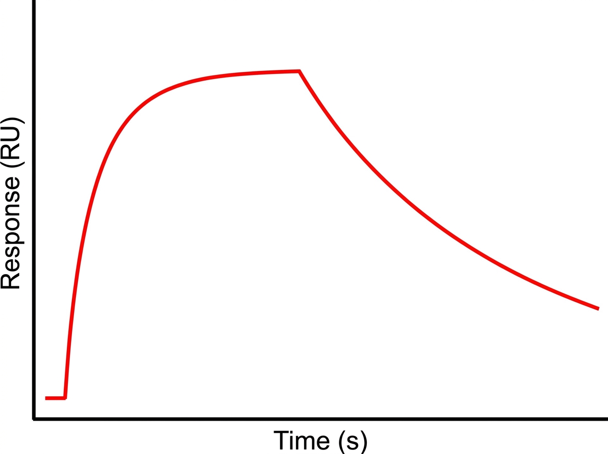
What is the technique shown in the graph that demonstrates antibody-antigen interaction and response kinetics?
Practice by Chapter
Spectrophotometry and Colorimetry
Practice Questions
Chromatography Techniques
Practice Questions
Electrophoresis and Applications
Practice Questions
Centrifugation and Ultracentrifugation
Practice Questions
Radioisotope Techniques
Practice Questions
Enzyme-Linked Immunosorbent Assay (ELISA)
Practice Questions
Polymerase Chain Reaction (PCR)
Practice Questions
Blotting Techniques: Southern, Northern, Western
Practice Questions
Mass Spectrometry in Biochemistry
Practice Questions
Recombinant DNA Technology
Practice Questions
DNA Sequencing
Practice Questions
Proteomics and Metabolomics
Practice Questions
Want unlimited practice?
Get full access to all questions, explanations, and performance tracking.
Scan to download app