
Respiratory and Airway Management — MCQs

On this page
A 72-year-old man has multiple injuries and an altered sensorium after a high-speed motor vehicle collision. He is intubated for his decreased mental status. During intubation, a large amount of gastric contents are noted in the posterior pharynx and he aspirates. What is the appropriate initial treatment?
Which statement is false regarding endotracheal tube cuffs?
If a satisfactory laryngeal view is not achieved during laryngoscopy, which maneuver is performed?
What is the approximate pressure that should be applied during cricoid pressure application?
Which supraglottic airway device offers the maximum airway seal during positive pressure ventilation?
Which of the following statements is true about the laryngeal mask airway?
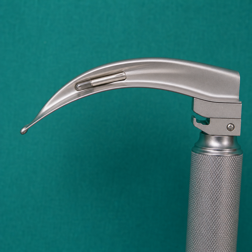
What is the name of the curved blade of a laryngoscope as depicted?
In a patient with Le Fort II, Le Fort III, and nasoethmoid fractures, what is the choice of intubation?
What is the landmark for superior laryngeal nerve block?
Which of the following conditions is NOT an indication for securing the airway?
Practice by Chapter
Respiratory Physiology
Practice Questions
Airway Anatomy
Practice Questions
Preoxygenation Techniques
Practice Questions
Mask Ventilation
Practice Questions
Supraglottic Airway Devices
Practice Questions
Direct Laryngoscopy
Practice Questions
Video Laryngoscopy
Practice Questions
Fiberoptic Intubation
Practice Questions
Surgical Airway Management
Practice Questions
One-Lung Ventilation Techniques
Practice Questions
Ventilation Strategies During Anesthesia
Practice Questions
Extubation Criteria and Techniques
Practice Questions
Want unlimited practice?
Get full access to all questions, explanations, and performance tracking.
Scan to download app