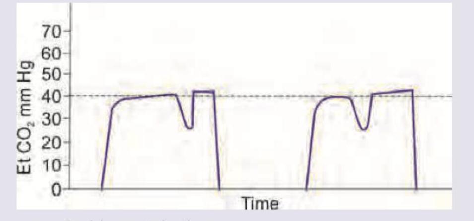
A patient is scheduled for emergency laparotomy for a perforated duodenal ulcer. He was administered 2 L of fluid and was wheeled into the OT. He was intubated and capnography recording shows the following recording. This indicates:

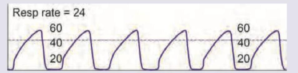
The capnography image shows:

What does the following capnographic recording represent?

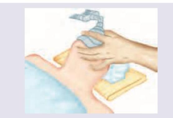
All are correct about airway management shown below except:

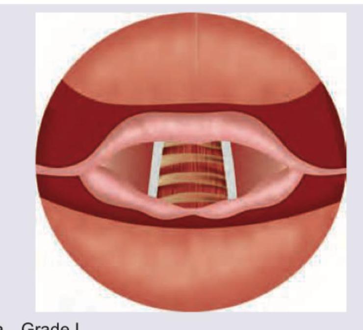
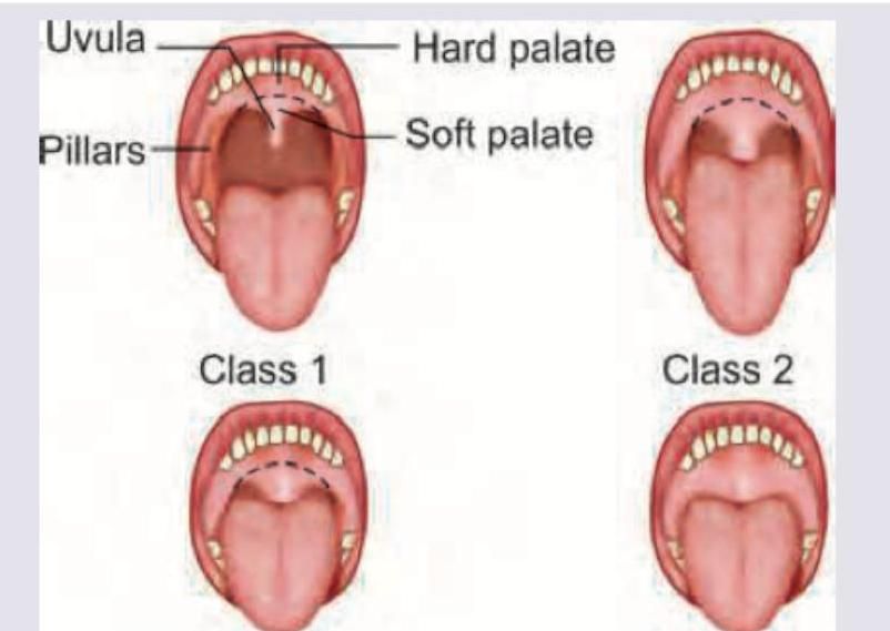
What is the grade of laryngeal view?

All are correct about the image shown except:

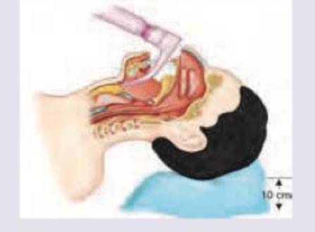
In correct positioning the tip of the instrument shown in the image should lie at:

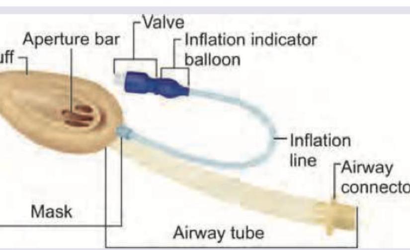
All are correct about the airway device shown below except: (Recent NEET Pattern 2016-17)

All are correct about the procedure being performed except: (Recent NEET Pattern 2016-17)

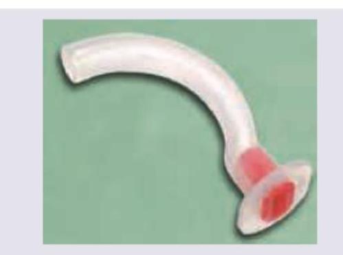
Which is correct about the instrument shown?

Explanation: ***Esophageal intubation*** - The capnography tracing shows **minimal and erratic CO2 waveforms**, which quickly fall to zero or near zero after a few breaths, indicating that the endotracheal tube is not in the trachea. - This pattern is characteristic of **esophageal intubation**, as there is little or no CO2 exhaled from the esophagus. *Placement of ET in right bronchus* - If the endotracheal tube is placed in the right bronchus (mainstem intubation), you would still see a **normal capnography waveform**, though often with signs of asymmetric chest rise and decreased breath sounds on the left side. - The problem would be related to **ventilation of only one lung**, not a complete absence of CO2. *Hypothermia* - While hypothermia can **decrease CO2 production** and lead to lower end-tidal CO2 values, it typically results in a **reduced but still discernible capnography waveform**, not a near absence of CO2. - The waveform morphology would remain largely normal, just shifted downwards. *Placement of ET in left bronchus* - Similar to right bronchus intubation, placement in the left bronchus would produce a **normal capnography waveform**, but with signs of unilateral lung ventilation (e.g., absent right-sided breath sounds). - The presence of a waveform indicates that the tube is in the airway, allowing for **gas exchange** and CO2 elimination from at least one lung.
Explanation: ***Curare cleft*** - The capnography waveform shows a **dip or cleft** during the expiratory plateau, which is characteristic of a 'curare cleft.' - This pattern occurs when the effect of **muscle relaxants (like curare)** is wearing off, and the patient attempts to breathe spontaneously, causing a transient decrease in CO2 exhalation. *Sudden extubation* - Sudden extubation would typically result in an **immediate and complete loss of the capnography waveform**, as there would be no CO2 reaching the sensor. - The image clearly displays repetitive, though altered, CO2 waveforms, indicating the patient is still intubated and ventilating. *Bronchospasm* - Bronchospasm is characterized by a **prolonged, upsloping phase three** (alpha angle) due to obstructed airflow, and often an **increased end-tidal CO2** (EtCO2) as CO2 clearance is impaired. - The waveform in the image does not show a prolonged upsloping phase or significantly increased EtCO2, but rather a distinct dip during the plateau. *Hypothermia* - Hypothermia generally causes a **decrease in EtCO2** due to reduced metabolic rate and CO2 production. - While it affects CO2 levels, it typically presents as a **lowered overall baseline** EtCO2, not a specific cleft within the expiratory plateau of individual breaths.
Explanation: ***Asthma*** - The capnographic tracing shows a characteristic **shark fin waveform**, indicative of **expiratory airflow obstruction**. - This waveform is generated due to uneven emptying of the alveoli, where CO2 continues to be exhaled at a reduced rate during the later phase of expiration. *Spontaneous extubation* - Spontaneous extubation would typically result in a **complete loss of the capnography waveform** due to disconnection from the airway. - The tracing shown still clearly depicts expired CO2, inconsistent with complete extubation. *Raised ICT* - Raised intracranial tension (ICT) can affect breathing patterns (e.g., Cheyne-Stokes, hyperventilation), but it does not directly produce a a **shark fin capnography waveform**. - Capnography reflects CO2 elimination, which can be indirectly affected by changes in ventilatory drive from raised ICT but not in this specific shape. *Air embolism* - An air embolism would cause a sudden **drop in end-tidal CO2 (EtCO2)**, often to zero, due to obstruction of pulmonary blood flow, leading to alveolar dead space. - The waveform shown does not depict a sudden drop to zero or significantly reduced EtCO2.
Explanation: *Index finger is placed on submental soft tissues to facilitate ventilation* - The image depicts a **face mask ventilation technique**. The index finger is typically placed on the **chin/mandible** to help secure the mask and perform a jaw thrust, not on the submental soft tissues in a way that directly facilitates ventilation. - Applying pressure to the submental soft tissues could potentially obstruct the airway rather than facilitate it. This statement describes an incorrect finger placement for effective mask ventilation. *Continued deflation of anesthesia reservoir bag indicates substantial leak around mask* - If the **anesthesia reservoir bag** continues to deflate during mask ventilation, it suggests that the delivered air is escaping, indicating a **poor seal** or substantial leak around the face mask. - A good mask seal is crucial for effective ventilation; continued deflation directly reflects a failure to maintain airway pressure. *Transparent mask allows observation of exhaled humidified gas and vomitus* - **Transparent face masks** are designed to allow practitioners to **visualize the patient's mouth and nose**. - This transparency enables quick assessment for signs of effective ventilation (e.g., fogging from **exhaled humidified gas**) and prompt detection of complications such as **vomitus** or secretions. *Face mask is contoured and conforms to varied facial features* - Modern **face masks** are designed with **malleable cushions** and ergonomic shapes to achieve an optimal seal on a wide range of patient facial anatomies. - This **contouring** is essential for minimizing leaks and ensuring efficient delivery of positive pressure ventilation.
Explanation: ***Grade I*** - In a **Grade I laryngeal view**, a **full view of the glottis** (vocal cords) is achieved during laryngoscopy. - This provides optimal conditions for endotracheal intubation, as seen in the image where the entire opening to the trachea is visible. *Grade II* - A **Grade II view** means only a **partial view of the glottis** is obtained, often with only the posterior commissure visible. - The anterior portion of the vocal cords may be obstructed by the epiglottis or other structures, making intubation more challenging. *Grade III* - **Grade III** indicates that only the **epiglottis** is visible, with no part of the glottis or vocal cords being seen. - Intubation is significantly more difficult in this scenario and often requires special techniques or adjuncts. *Grade IV* - A **Grade IV view** is the most difficult, where **neither the epiglottis nor the glottis** can be visualized. - This implies that only the soft palate or base of the tongue is seen, representing a very challenging airway.
Explanation: ***Not influenced by tongue mobility and size*** - The **Mallampati classification** is significantly influenced by **tongue size and mobility**, as a large or non-mobile tongue can obstruct the view of the posterior pharyngeal structures. - The classification assesses the visible structures of the soft palate, uvula, and tonsillar pillars, which can be obscured by the tongue. *Mallampati classification* - The image clearly depicts the structures observed in the **Mallampati classification** system (uvula, pillars, hard palate, soft palate, and progressively less visibility in classes 1-4). - This classification is a widely used tool for assessing airway patency. *Helps in identifying cases with difficult orotracheal intubation* - The **Mallampati classification** is a primary bedside tool used to predict the difficulty of **orotracheal intubation**. - Higher Mallampati classes (3 and 4) are associated with a greater likelihood of difficult intubation due to reduced visibility of the pharyngeal structures. *Has limited utility in patients with reduced neck extension* - Poor **neck extension** can limit the alignment of the oral, pharyngeal, and laryngeal axes, making direct laryngoscopy and intubation difficult. - The Mallampati classification primarily assesses oral cavity visualization and does not account for limitations imposed by neck mobility, thereby having reduced predictive power in such cases.
Explanation: ***Epiglottis*** - The image shows a **Laryngeal Mask Airway (LMA)**, which is designed to sit in the hypopharynx, with its tip resting at the **epiglottis**. - This positioning allows the LMA to create a seal around the laryngeal inlet, facilitating effective ventilation without entering the trachea. *Vocal cords* - The LMA is designed to provide a seal *above* the vocal cords, ensuring ventilation of the trachea without direct intubation of the vocal cords themselves. - Positioning the tip *at* the vocal cords would hinder proper airway sealing and could cause trauma. *Thyroid cartilage* - The thyroid cartilage is an anterior neck structure and is not the anatomical landmark for the tip of a properly placed LMA. - The LMA sits deeper in the pharynx, above the glottic opening, making the epiglottis the relevant landmark. *Above esophagus* - While the LMA sits **above the esophageal inlet**, diverting air primarily into the trachea, its *tip* specifically rests at the epiglottis, covering the laryngeal opening. - Stating "above the esophagus" is too general; the precise anatomical placement for the tip is at the epiglottis.
Explanation: ***It should remain in place till airway reflexes are regained*** - The device shown is a **Laryngeal Mask Airway (LMA)**, which is typically removed once the patient shows signs of **regaining airway reflexes**, such as gagging or coughing. - Keeping it in place too long after airway reflexes return increases the risk of **laryngospasm** and patient discomfort. *Contraindicated in patients with pharyngeal abscess* - The LMA is **contraindicated** in patients with a pharyngeal abscess due to the risk of **rupturing the abscess** and causing aspiration or spreading infection. - This is a valid contraindication, so the statement is correct in its assertion. *Protects the larynx from gastric contents* - While an LMA provides **some protection** against aspiration by sealing off the larynx from the pharynx, it does not offer the same level of definitive protection as a cuffed **endotracheal tube**. - There is still a risk of **regurgitation and aspiration** of gastric contents, especially in patients with high risk factors. *Helps in passage of tracheal tube in patient with difficult airway* - The LMA can indeed be used as a conduit for **fiberoptic intubation** or with specialized catheters to facilitate the placement of an endotracheal tube in cases of a **difficult airway**. - This is a recognized use of the LMA in airway management algorithms.
Explanation: ***Tip of curved blade is inserted into aryepiglottic fold*** - This statement is incorrect because the tip of a **Macintosh blade** (curved blade) is designed to be placed in the **vallecula**, the space between the base of the tongue and the epiglottis, not the aryepiglottic fold. - Positioning in the vallecula allows the blade to indirectly lift the **epiglottis**, exposing the vocal cords. *Macintosh laryngoscope is being used for intubation* - The image clearly shows a **curved laryngoscope blade**, which is characteristic of the **Macintosh blade**. - The Macintosh laryngoscope is commonly used for **oral endotracheal intubation** to visualize the vocal cords. *Tongue swept to left using flange of blade* - During direct laryngoscopy, the laryngoscope blade is inserted on the **right side of the tongue** and then used to sweep the tongue to the left. - This maneuver helps to clear the line of sight and prevent obstruction from the tongue. *Handle raised up and away, perpendicular to patient's mandible to expose vocal cords* - To properly expose the vocal cords, the laryngoscope handle should be lifted **upward and outward** along the axis of the handle, away from the patient's face. - This action elevates the **epiglottis** and associated structures, providing a clear view of the **larynx**.
Explanation: ***Insert with rotation movement of 90 degrees with convex upwards*** - The image shows an **oropharyngeal airway (OPA)**. The correct insertion technique involves inserting it with the **convex side upwards** and rotating it **90 degrees** as it reaches the soft palate. - This method prevents pushing the tongue backward and ensures proper positioning with the **concave side** facing the tongue to maintain airway patency. *Insert in clonus stage to prevent tongue bite* - Inserting an OPA during active **clonus stage** is dangerous and contraindicated due to risk of injury and **vomiting**. - OPAs should be inserted only when the patient is **deeply unconscious** and not actively seizing. *Assist in oropharyngeal suctioning* - While an OPA creates a clear pathway, its primary function is **airway maintenance**, not suctioning assistance. - **Suctioning** can be performed with or without an OPA, but the device doesn't actively assist the process. *Can prevent aspiration of GI contents* - An OPA **does not protect** against aspiration as it sits in the oropharynx without sealing the trachea. - Only **endotracheal intubation** with cuff inflation can effectively prevent aspiration of gastric contents.
Respiratory Physiology
Practice Questions
Airway Anatomy
Practice Questions
Preoxygenation Techniques
Practice Questions
Mask Ventilation
Practice Questions
Supraglottic Airway Devices
Practice Questions
Direct Laryngoscopy
Practice Questions
Video Laryngoscopy
Practice Questions
Fiberoptic Intubation
Practice Questions
Surgical Airway Management
Practice Questions
One-Lung Ventilation Techniques
Practice Questions
Ventilation Strategies During Anesthesia
Practice Questions
Extubation Criteria and Techniques
Practice Questions
Get full access to all questions, explanations, and performance tracking.
Start For Free