
Respiratory and Airway Management — MCQs

On this page
In controlled ventilation, which of the following statements is true?
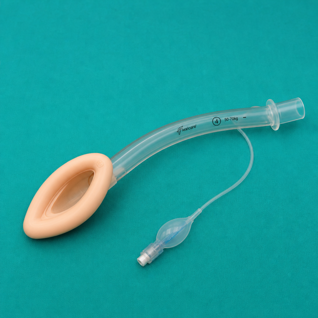
Identify the airway device shown in the image.
Size of LMA for a 15kg child is?
What is Mallampati classification used for?
Which of the following is NOT a contraindication for bag and mask ventilation?
What is the primary use of the Macintosh laryngoscope?
The laryngeal mask airway is used for securing the airway in all of the following conditions, EXCEPT:
In which of the following scenarios is the laryngeal mask airway (LMA) typically not used?
What is the standard method used to differentiate between endotracheal and esophageal intubation?
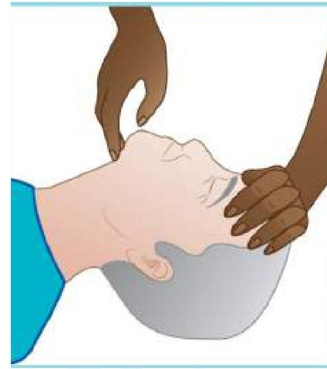
Identify the maneuver being performed.
Practice by Chapter
Respiratory Physiology
Practice Questions
Airway Anatomy
Practice Questions
Preoxygenation Techniques
Practice Questions
Mask Ventilation
Practice Questions
Supraglottic Airway Devices
Practice Questions
Direct Laryngoscopy
Practice Questions
Video Laryngoscopy
Practice Questions
Fiberoptic Intubation
Practice Questions
Surgical Airway Management
Practice Questions
One-Lung Ventilation Techniques
Practice Questions
Ventilation Strategies During Anesthesia
Practice Questions
Extubation Criteria and Techniques
Practice Questions
Want unlimited practice?
Get full access to all questions, explanations, and performance tracking.
Scan to download app