
Respiratory and Airway Management — MCQs

On this page
Endotracheal tube in the esophagus is best assessed by:
During rapid sequence intubation in a child after taking brief history and clinical examination next step is:
Effective adjuvant in attenuating hypertension and tachycardia associated with laryngoscopy and intubation?
A patient is admitted following a road traffic accident. He has sustained significant blunt injury to his head, chest and abdomen and has a Glasgow Coma Scale score of 8/15. His saturations are poor at 89% on 15 L of oxygen a rebreathing mask. You note bruising around both eyes and blood-stained fluid issuing from his left ear, which forms concentric circles when dripped on a white sheet. You wish to support his airway to improve oxygenation. The first choice of airway adjunct would be
On doing laparoscopic cholecystectomy patient developed wheezing. Which of the following is used in the treatment?
Helium is used along with oxygen instead of plain oxygen because
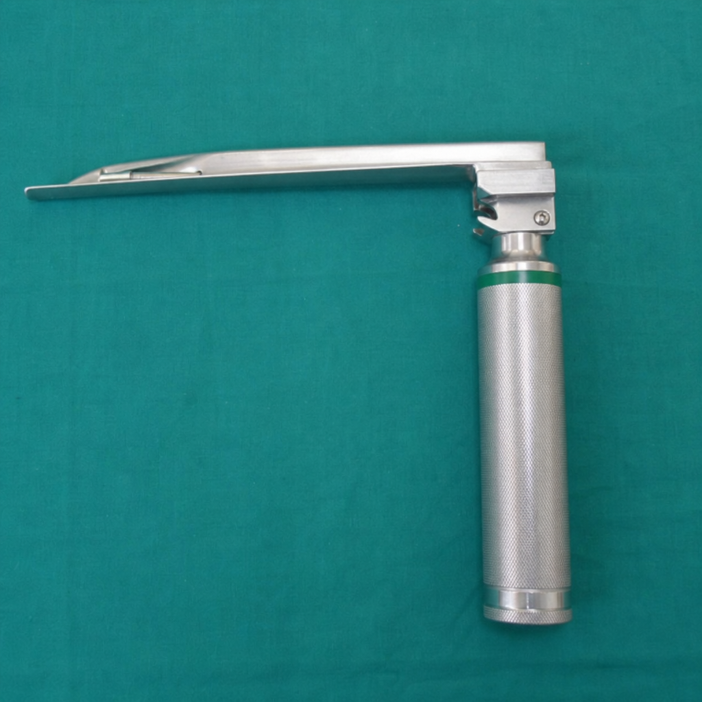
WHAT IS THE TYPE OF LARYNGOSCOPE SHOWN BELOW ?
Which of the following is the most common postoperative complication related to intubation:
Intraocular pressure rises in ?
The Blade of the laryngoscope used in intubation of newborn is
Practice by Chapter
Respiratory Physiology
Practice Questions
Airway Anatomy
Practice Questions
Preoxygenation Techniques
Practice Questions
Mask Ventilation
Practice Questions
Supraglottic Airway Devices
Practice Questions
Direct Laryngoscopy
Practice Questions
Video Laryngoscopy
Practice Questions
Fiberoptic Intubation
Practice Questions
Surgical Airway Management
Practice Questions
One-Lung Ventilation Techniques
Practice Questions
Ventilation Strategies During Anesthesia
Practice Questions
Extubation Criteria and Techniques
Practice Questions
Want unlimited practice?
Get full access to all questions, explanations, and performance tracking.
Scan to download app