
Anesthetic Equipment and Monitoring — MCQs

On this page
Which of the following is the most common method used to know depth of anaesthesia?
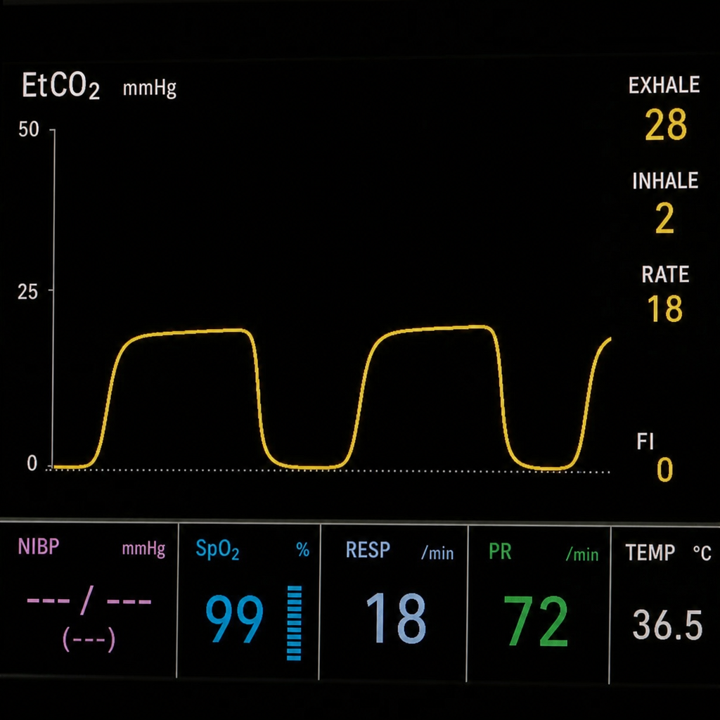
What does the image of the Capnograph depict?
Murphy's eye is seen in -
What is the primary use of the Mapleson E anesthesia circuit?
What is the primary purpose of the Pin Index System in anesthesia machines?
What does the acronym DISS stand for in the context of medical gas supply?
Which gas is commonly used in the medical field for rapid inflation of devices such as air embolism balloons?
What is the pressure at which oxygen is stored?
Which of the following systems is specifically designed to produce Positive End-Expiratory Pressure (PEEP) in mechanical ventilation?
What does the image of the Capnograph depict?

Practice by Chapter
Anesthesia Machine Components
Practice Questions
Breathing Systems
Practice Questions
Vaporizers
Practice Questions
Gas Cylinders and Pipeline Supply
Practice Questions
Anesthesia Ventilators
Practice Questions
Standard Monitoring: ECG, BP, Pulse Oximetry
Practice Questions
Capnography
Practice Questions
Neuromuscular Monitoring
Practice Questions
Temperature Monitoring
Practice Questions
Invasive Hemodynamic Monitoring
Practice Questions
Equipment Troubleshooting
Practice Questions
Safety Features in Modern Anesthesia Equipment
Practice Questions
Want unlimited practice?
Get full access to all questions, explanations, and performance tracking.
Scan to download app