
Anesthetic Equipment and Monitoring — MCQs

On this page
In medical applications, the best fuel gas when used with oxygen is:
All of the following cause a decrease in pulse oximeter readings except:
In anesthesia, fitting of wrong gas cylinder to the anesthesia machine can be prevented by:
Which of the following is NOT a rebreathing system used in anesthesia?
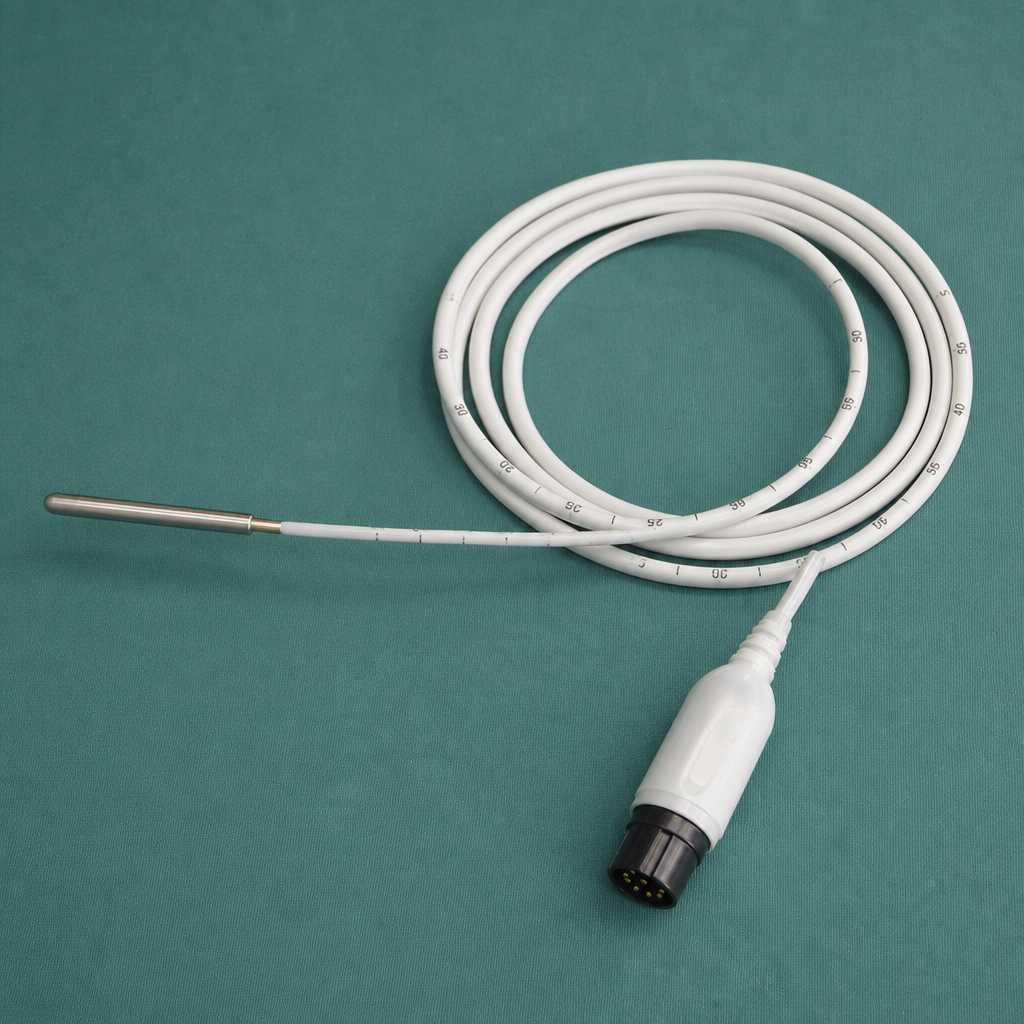
The instrument below is used for
A 40–year female has to undergo incisional hernia surgery under general anaesthesia. She complains of awareness during her past cesarean section. Which of the following monitoring techniques can be used to prevent such awareness ?
Which nerve is used for monitoring anesthesia during surgery?
Which is wrong regarding somato sensory evoked potentials (SSEP)?
Gas stored in liquid form is:
True about anaesthesia breathing circuit is
Practice by Chapter
Anesthesia Machine Components
Practice Questions
Breathing Systems
Practice Questions
Vaporizers
Practice Questions
Gas Cylinders and Pipeline Supply
Practice Questions
Anesthesia Ventilators
Practice Questions
Standard Monitoring: ECG, BP, Pulse Oximetry
Practice Questions
Capnography
Practice Questions
Neuromuscular Monitoring
Practice Questions
Temperature Monitoring
Practice Questions
Invasive Hemodynamic Monitoring
Practice Questions
Equipment Troubleshooting
Practice Questions
Safety Features in Modern Anesthesia Equipment
Practice Questions
Want unlimited practice?
Get full access to all questions, explanations, and performance tracking.
Scan to download app