
Upper Limb — MCQs

On this page
Which of the following is not intracapsular -
Serratus anterior is supplied by which nerve?
Middle palmar space ends distally:
A cut injury on wrist causes the inability of thumb to touch the tip of little finger, the nerve likely to be damaged is -
Pronator teres syndrome is related to which nerve?
Which of the following is not a flexor of the forearm?
Clinical testing of the function of the long thoracic nerve is done by:
What are the attachments of the ulnar collateral ligament?
Power grip of hand depends on?
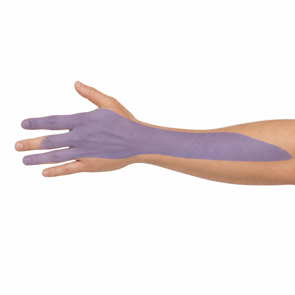
Which of the following nerves has the shaded cutaneous territory
Practice by Chapter
Pectoral Region and Axilla
Practice Questions
Arm and Cubital Fossa
Practice Questions
Forearm and Hand
Practice Questions
Joints of Upper Limb
Practice Questions
Nerves of Upper Limb
Practice Questions
Arterial Supply and Venous Drainage
Practice Questions
Lymphatic Drainage
Practice Questions
Muscles and Their Actions
Practice Questions
Applied Anatomy and Clinical Correlations
Practice Questions
Surface Anatomy and Landmarks
Practice Questions
Want unlimited practice?
Get full access to all questions, explanations, and performance tracking.
Scan to download app