
Upper Limb — MCQs

On this page
NOT a content of carpal tunnel:-
Retraction of scapula is done by
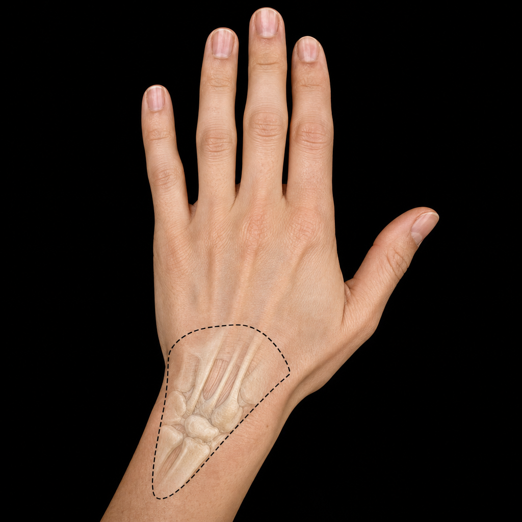
An injury to the shown area can lead to fracture of which bone
Erb-Duchenne paralysis occurs by lesion to brachial plexus at the level of
In a diving accident that severed the spinal cord below the sixth cervical vertebra, which of the following muscles would be affected?
Extensor carpi radialis longus is
In interphalangeal joint, the capsule is thinnest on the -
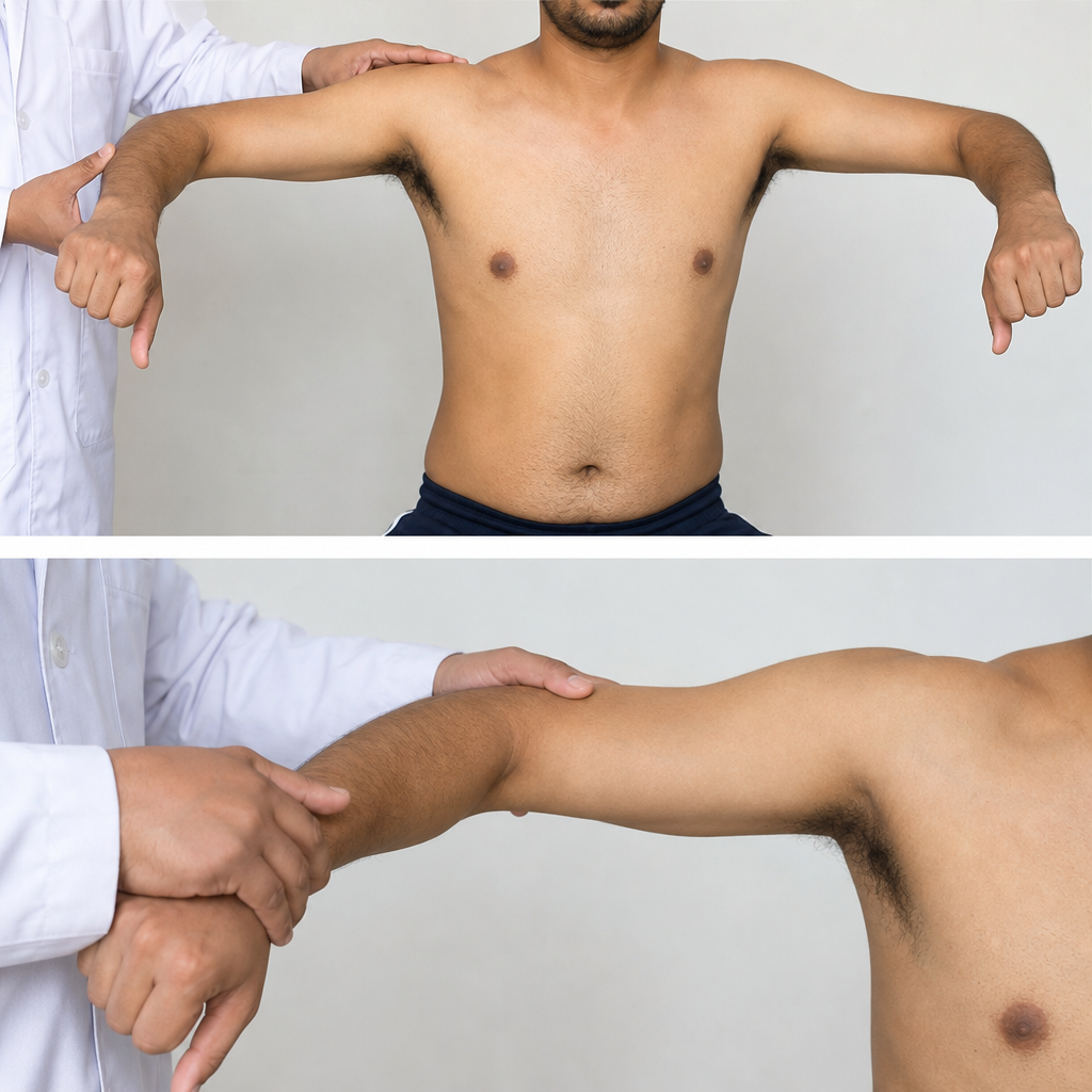
Test shown in the video is for which structure & its nerve supply:
Partial claw hand is caused by lesion involving the:
In cubital fossa, which structure is the most medial
Practice by Chapter
Pectoral Region and Axilla
Practice Questions
Arm and Cubital Fossa
Practice Questions
Forearm and Hand
Practice Questions
Joints of Upper Limb
Practice Questions
Nerves of Upper Limb
Practice Questions
Arterial Supply and Venous Drainage
Practice Questions
Lymphatic Drainage
Practice Questions
Muscles and Their Actions
Practice Questions
Applied Anatomy and Clinical Correlations
Practice Questions
Surface Anatomy and Landmarks
Practice Questions
Want unlimited practice?
Get full access to all questions, explanations, and performance tracking.
Scan to download app