
Upper Limb — MCQs

On this page
A patient who has taken the first COVID vaccine comes for the second dose. An astute nurse noticed that the shoulder was flabby, flat, and was asymmetrical. There was an associated loss of contour of the shoulder joint. Injury to which of the structures might have resulted and was avoidable?
A patient presents with difficulty extending their wrist following trauma to the posterior forearm. Which of the following muscles would be most affected by injury to the posterior interosseous nerve?
A 45-year-old woman undergoes a modified radical mastectomy for breast cancer. Following the procedure, she experiences numbness in the medial aspect of her upper arm. Which of the following nerves was most likely injured during the surgery?
A 16-year-old boy is brought to the emergency department after being tackled at a football game. Per his mom, he is the quarterback of his team and was head-butted in the left shoulder region by the opposing team. Shortly after, the mother noticed that his left arm was hanging by his torso and his hand was “bent backwards and facing the sky.” The patient denies head trauma, loss of consciousness, sensory changes, or gross bleeding. A physical examination demonstrates weakness in abduction, lateral rotation, flexion, and supination of the left arm and tenderness of the left shoulder region with moderate bruising. Radiograph of the left shoulder and arm is unremarkable. Which of the following is most likely damaged in this patient?
Tendons in the 2nd compartment of wrist?
Which bone connects the sternum to the scapula?
Which of the following structures passes through Guyon's canal?
Pointing index finger is seen in which nerve injury
Winging of scapula is due to paralysis of
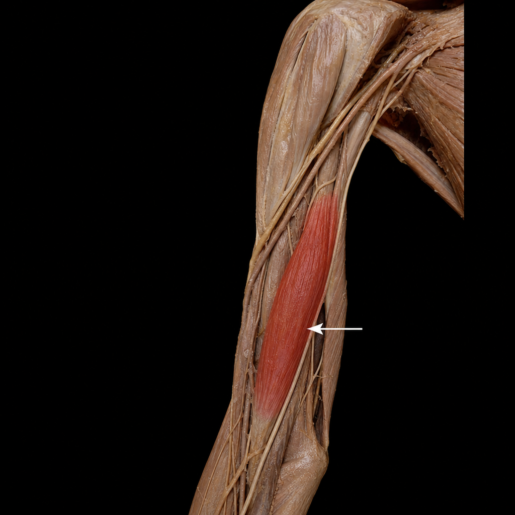
The highlighted muscle is supplied by which nerve?
Practice by Chapter
Pectoral Region and Axilla
Practice Questions
Arm and Cubital Fossa
Practice Questions
Forearm and Hand
Practice Questions
Joints of Upper Limb
Practice Questions
Nerves of Upper Limb
Practice Questions
Arterial Supply and Venous Drainage
Practice Questions
Lymphatic Drainage
Practice Questions
Muscles and Their Actions
Practice Questions
Applied Anatomy and Clinical Correlations
Practice Questions
Surface Anatomy and Landmarks
Practice Questions
Want unlimited practice?
Get full access to all questions, explanations, and performance tracking.
Scan to download app