The midpalmar space of the hand communicates with all the following structures except?
In Erb-Duchene paralysis, the injury is limited to which spinal nerve roots?
Which type of epiphysis is the coracoid process?
All of the following are features of musculocutaneous nerve injury at the axilla, except?
An isolated avulsion fracture of the lesser tubercle of the humerus in a 16-year-old athlete is caused by the forceful contraction of which of the following muscles?
A fall on an outstretched hand can lead to a fracture of which of the following bones?
What is the nerve supply of the Trapezius muscle?
Which of the following is a subcutaneous muscle?
Damage to the floor of the anatomic snuffbox and origin of the abductor pollicis brevis most likely involves which of the following bones?
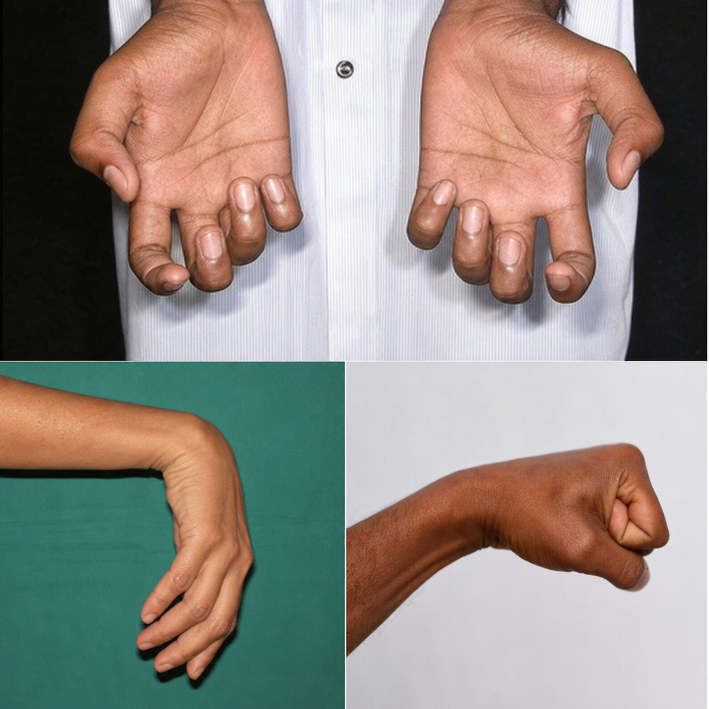
Which of the following hand deformities is presented?

Explanation: **Explanation:** The **midpalmar space** is a deep fascial space of the hand located medial to the intermediate palmar septum [1]. Understanding its boundaries and communications is crucial for predicting the spread of hand infections. **1. Why Option B is correct:** The midpalmar space communicates distally with the **fascial sheaths of the 2nd, 3rd, and 4th lumbrical muscles** (which pass through the 2nd, 3rd, and 4th web spaces). The **1st lumbrical** is located in the **thenar space**, not the midpalmar space. Therefore, an infection in the midpalmar space will not directly track into the 1st lumbrical canal. **2. Why the other options are incorrect:** * **Forearm space (Space of Parona):** Proximally, the midpalmar space is continuous with the forearm space deep to the flexor tendons via the carpal tunnel. * **Fascial sheaths of 2nd and 3rd lumbricals:** These canals serve as the primary distal drainage/communication routes for the midpalmar space. The 2nd lumbrical corresponds to the index finger, and the 3rd to the middle finger. **High-Yield Clinical Pearls for NEET-PG:** * **Boundaries:** The midpalmar space is separated from the thenar space by a fibrous septum attached to the **3rd metacarpal bone** [1]. * **Clinical Presentation:** Infections here result in the loss of the normal concavity of the palm ("ballooning" of the palm) [1]. * **Kanavel’s Signs:** While primarily for tenosynovitis, remember that midpalmar space infections often occur secondary to neglected tenosynovitis of the middle, ring, or little fingers. * **The "U-Bursa" Communication:** The synovial sheath of the little finger is usually continuous with the common flexor sheath (ulnar bursa), which lies in the midpalmar space.
Explanation: Erb-Duchenne Paralysis (or Erb’s Palsy) is a clinical condition resulting from an injury to the upper trunk of the brachial plexus. The upper trunk is formed by the union of the anterior primary rami of the C5 and C6 spinal nerves. 1. Why Option D is Correct: The site of injury is typically Erb’s point, where six nerves meet. A forceful increase in the angle between the head and shoulder (e.g., birth trauma or falling on the shoulder) stretches or tears the C5 and C6 roots [1]. This leads to paralysis of muscles supplied by these segments, most notably the deltoid, biceps brachii, brachialis, and supinator. 2. Why Other Options are Incorrect: * Options A & B (C2-C4): These roots contribute to the cervical plexus, which supplies the skin and muscles of the neck and the diaphragm (via the phrenic nerve, C3-C5). They are not part of the brachial plexus. * Option C (C4-C5): While C5 is involved, C4 only provides a small "pre-fixed" contribution to the plexus. The hallmark of Erb’s palsy specifically involves the C6 root. Clinical Pearls for NEET-PG: * Deformity: The classic clinical presentation is the "Policeman’s tip hand" or "Waiter’s tip hand." The arm hangs by the side, is adducted (loss of abductors), medially rotated (loss of lateral rotators), and the forearm is extended and pronated [1]. * Nerves Involved: Suprascapular nerve, nerve to subclavius, and the musculocutaneous and axillary nerves are severely affected. * Contrast with Klumpke’s Paralysis: Klumpke’s involves the lower trunk (C8-T1), resulting in a "claw hand" deformity due to the loss of intrinsic hand muscles.
Explanation: ### Explanation The **coracoid process** of the scapula is a classic example of an **atavistic epiphysis**. **1. Why Atavistic Epiphysis is Correct:** An atavistic epiphysis represents a bone that was phylogenetically independent in lower animals (ancestors) but has become fused to another bone in humans during evolution. In lower vertebrates (like reptiles and birds), the coracoid is a separate bone of the pectoral girdle that connects the scapula to the sternum. In humans, it has lost its independent function and exists only as a process that fuses with the scapula. **2. Analysis of Incorrect Options:** * **Pressure Epiphysis (Option A):** These are found at the ends of long bones and are subjected to pressure during weight-bearing or joint movement (e.g., Head of the femur, Lower end of the radius). They contribute to the length of the bone [1]. * **Traction Epiphysis (Option B):** These develop due to the "tug" or pull of attached tendons or muscles. They do not take part in joint formation (e.g., Greater and lesser trochanters of the femur, Medial and lateral epicondyles of the humerus). * **Aberrant Epiphysis (Option C):** These are deviations from the norm and are not always present (e.g., Epiphysis at the head of the first metacarpal or the base of other metacarpals). **3. NEET-PG High-Yield Pearls:** * **Other Atavistic Examples:** The **Os trigonum** (posterior tubercle of the talus) and the **tubercle of the ischium** (in some classifications). * **Coracoid Ossification:** It develops from two centers—a primary center for the main body and a secondary center (subcoracoid) for the base. * **Clinical Significance:** The coracoid process serves as the "Lighthouse of the Shoulder" for surgeons and is the attachment site for the "Short head of Biceps," "Coracobrachialis," and "Pectoralis minor" muscles.
Explanation: ### Explanation The **Musculocutaneous nerve (C5–C7)** is a branch of the lateral cord of the brachial plexus. It supplies the muscles of the anterior compartment of the arm: **Coracobrachialis, Biceps Brachii, and Brachialis.** **1. Why "Loss of flexion of the shoulder" is the correct answer (EXCEPT):** While the Biceps brachii and Coracobrachialis do assist in shoulder flexion, they are only **accessory flexors**. The primary (chief) flexors of the shoulder are the **Deltoid (anterior fibers)** and **Pectoralis major (clavicular head)**, which are supplied by the Axillary and Lateral Pectoral nerves, respectively. Therefore, even with a musculocutaneous nerve injury, shoulder flexion is significantly weakened but **not lost**. **2. Analysis of Incorrect Options:** * **Loss of flexion at the elbow:** The Biceps brachii and Brachialis are the primary flexors of the elbow. Their paralysis leads to a profound loss of elbow flexion. * **Loss of supination of the forearm:** The Biceps brachii is the **most powerful supinator** of the forearm when the elbow is flexed. Injury results in a major loss of supinatory power (though the Supinator muscle, supplied by the radial nerve, remains intact). * **Loss of sensation on the radial side of the forearm:** After supplying the muscles, the nerve continues as the **Lateral Cutaneous Nerve of the Forearm**, providing sensation to the radial (lateral) aspect of the forearm. **Clinical Pearls for NEET-PG:** * **Testing:** The integrity of the musculocutaneous nerve is clinically tested via the **Biceps Reflex (C5-C6)**. * **Sensory Loss:** The sensory deficit is limited to the forearm; there is **no sensory loss in the arm** because the medial and lateral cutaneous nerves of the arm arise directly from the brachial plexus cords. * **Vulnerability:** It is most commonly injured by heavy pressure in the axilla or during shoulder dislocations.
Explanation: The correct answer is **Subscapularis**. This question tests your knowledge of functional anatomy and muscle insertions on the proximal humerus. **1. Why Subscapularis is correct:** The **lesser tubercle** of the humerus serves as the specific insertion point for only one rotator cuff muscle: the **Subscapularis**. An avulsion fracture occurs when a tendon or ligament pulls a fragment of bone away from the main body. In young athletes, forceful external rotation or sudden contraction of the subscapularis (often during sports like wrestling or throwing) can lead to an isolated avulsion of the lesser tubercle. **2. Why the other options are incorrect:** * **Supraspinatus, Infraspinatus, and Teres minor:** These three muscles collectively insert onto the **greater tubercle** of the humerus. * **Supraspinatus:** Inserts on the superior impression. * **Infraspinatus:** Inserts on the middle impression. * **Teres minor:** Inserts on the inferior impression. An avulsion of the greater tubercle would involve one or more of these muscles, not the subscapularis. **3. Clinical Pearls & High-Yield Facts for NEET-PG:** * **Rotator Cuff Mnemonic (SITS):** **S**upraspinatus, **I**nfraspinatus, **T**eres minor (all Greater Tubercle) and **S**ubscapularis (Lesser Tubercle). * **Function:** The Subscapularis is the primary **internal rotator** of the humerus. * **Nerve Supply:** It is supplied by the **Upper and Lower Subscapular nerves** (C5, C6). * **Clinical Test:** The **Lift-off test** or **Belly-press test** is used to assess the integrity of the subscapularis muscle/tendon. * **Bicipital Groove:** Located between the greater and lesser tubercles, it lodges the long head of the biceps brachii tendon. The **Latissimus dorsi** inserts into the floor of this groove.
Explanation: Explanation: A **Fall on an Outstretched Hand (FOOSH)** is a classic mechanism of injury in orthopedics where force is transmitted proximally from the palm through the carpus, forearm, and shoulder girdle. 1. **Why Option A is Correct:** * **Scaphoid:** It is the most commonly fractured carpal bone [1]. During FOOSH, the scaphoid is compressed between the radius and the distal carpal row (specifically the capitate), leading to a fracture, usually at the waist [1]. * **Capitate:** While less common than the scaphoid, the capitate is the largest carpal bone and lies in the direct line of force transmission from the third metacarpal to the radius. * **Clavicle:** The force travels up the limb through the radius, humerus, and glenoid. The clavicle acts as a strut connecting the upper limb to the axial skeleton; its weakest point (junction of medial 2/3 and lateral 1/3) often fails under this transmitted longitudinal stress. 2. **Why other options are incorrect:** * **Option B:** While correct, it is incomplete compared to Option A. * **Option C:** FOOSH typically causes fractures of the distal radius (Colles’ fracture) rather than the head of the ulna. Ulnar injuries are more common in direct trauma (e.g., Nightstick fracture). * **Option D:** A fracture of the radial styloid process (Chauffeur’s fracture) usually results from direct compression or avulsion, not the generalized longitudinal force transmission seen in a standard FOOSH. **High-Yield Clinical Pearls for NEET-PG:** * **Scaphoid Fracture:** Look for tenderness in the **Anatomical Snuffbox**. The most serious complication is **Avascular Necrosis (AVN)** due to retrograde blood supply [1]. * **Colles’ Fracture:** Distal radius fracture with **posterior (dorsal) displacement**, resulting in a "Dinner Fork Deformity." * **Smith’s Fracture:** Reverse Colles’ (ventral displacement) caused by a fall on a flexed wrist. * **Clavicle:** The most common site of fracture is the mid-shaft.
Explanation: The **Trapezius** is a large, diamond-shaped muscle of the back and neck. Its nerve supply is unique and a frequent high-yield topic in NEET-PG: **1. Why Spinal Accessory Nerve is Correct:** The Trapezius receives its **motor supply** from the **Spinal Accessory Nerve (CN XI)**. This nerve originates from the upper five or six cervical segments of the spinal cord, enters the cranium via the foramen magnum, and exits through the jugular foramen to supply the Sternocleidomastoid and Trapezius. Additionally, the muscle receives **sensory (proprioceptive) fibers** from the ventral rami of **C3 and C4** spinal nerves. **2. Why Incorrect Options are Wrong:** * **Hypoglossal nerve (CN XII):** Supplies all intrinsic and extrinsic muscles of the tongue (except Palatoglossus). * **Trochlear nerve (CN IV):** A pure motor nerve supplying only the Superior Oblique muscle of the eye. * **Trigeminal nerve (CN V):** Primarily supplies the muscles of mastication (via the mandibular branch) and provides facial sensation. **3. Clinical Pearls for NEET-PG:** * **Testing:** To test the Trapezius, ask the patient to "shrug their shoulders" against resistance. * **Injury:** Damage to CN XI (often during lymph node biopsy in the posterior triangle) results in drooping of the shoulder and an inability to abduct the arm above 90 degrees (due to loss of scapular rotation). * **Dual Supply:** Remember that while CN XI is motor, C3-C4 are sensory. This distinction is crucial for exams.
Explanation: The correct answer is **Palmaris brevis**. In anatomy, a **subcutaneous muscle** (or muscle of the panniculus carnosus) is one that is located within the superficial fascia and inserts directly into the skin rather than onto bone [1]. * **Palmaris Brevis:** This is a thin, quadrilateral muscle located in the superficial fascia of the hypothenar eminence [1]. It originates from the palmar aponeurosis and flexor retinaculum and inserts into the **dermis of the skin** on the ulnar border of the hand. Its primary function is to wrinkle the skin of the hypothenar eminence and deepen the hollow of the palm, improving grip [1]. **Analysis of Incorrect Options:** * **Sternocleidomastoid:** A major muscle of the neck that lies deep to the platysma. It originates from the sternum/clavicle and inserts into the mastoid process (bone). * **Mylohyoid:** A suprahyoid muscle forming the floor of the oral cavity; it attaches to the mylohyoid line of the mandible. * **Palmaris Longus:** A vestigial muscle of the forearm. While its tendon is superficial, it is a skeletal muscle that inserts into the palmar aponeurosis, not the skin. **Clinical Pearls for NEET-PG:** 1. **Ulnar Nerve:** The palmaris brevis is unique because it is the **only** muscle supplied by the **superficial branch of the ulnar nerve** [1]. 2. **Protection:** It serves to protect the underlying ulnar artery and nerve from pressure during gripping. 3. **Other Subcutaneous Muscles:** Other examples include the **Platysma** (neck) and the **muscles of facial expression**.
Explanation: ### Explanation The correct answer is **Trapezium (Option B)**. This question tests your knowledge of the bony landmarks of the wrist and the origins of the intrinsic muscles of the hand. **1. Why Trapezium is Correct:** The **anatomic snuffbox** is a triangular depression on the radial aspect of the wrist. Its floor is formed by two bones: the **Scaphoid** (proximally) and the **Trapezium** (distally) [1]. Furthermore, the **Abductor Pollicis Brevis (APB)**—a member of the thenar muscle group—takes its origin from the tubercle of the trapezium and the associated flexor retinaculum [1]. Therefore, a bone that contributes to both the floor of the snuffbox and the origin of the APB must be the trapezium. **2. Why Other Options are Incorrect:** * **Scaphoid (Option A):** While the scaphoid forms the proximal part of the snuffbox floor and is the most commonly fractured carpal bone [1], it is **not** the origin for the abductor pollicis brevis. * **Lunate (Option C):** The lunate is located in the proximal row of carpal bones but is situated medially to the scaphoid. It does not form the snuffbox floor nor serve as an origin for thenar muscles. * **Capitate (Option D):** The capitate is the largest carpal bone, located centrally in the distal row. It does not contribute to the radial-sided anatomic snuffbox. **Clinical Pearls for NEET-PG:** * **Snuffbox Boundaries:** Lateral (Abductor pollicis longus & Extensor pollicis brevis); Medial (Extensor pollicis longus) [1]. * **Contents:** The **Radial Artery** passes through the snuffbox. * **Tenderness:** Tenderness in the snuffbox classically indicates a **Scaphoid fracture**, which carries a high risk of avascular necrosis (AVN) due to retrograde blood supply [1]. * **Thenar Muscles (Meat):** Remember the mnemonic **OAF** (Opponens pollicis, Abductor pollicis brevis, Flexor pollicis brevis)—all primarily originate from the trapezium and flexor retinaculum [1].
Explanation: ***All of the above*** - The hand deformity shows features consistent with **combined nerve injuries**, including **clawing** (ulnar), **thenar wasting** (median), and **intrinsic muscle paralysis** affecting both nerve territories. - **Klumpke's paralysis** (C8-T1 root lesion) causes loss of all **intrinsic hand muscles**, resulting in combined ulnar and median nerve territory deficits. *Ulnar nerve injury* - Would cause **clawing deformity** and **hypothenar wasting**, but **thenar muscles** would remain intact. - Does not explain the **complete intrinsic muscle paralysis** seen in this presentation. *Median nerve injury* - Would cause **thenar wasting** and loss of **opposition**, but **intrinsic muscles** supplied by ulnar nerve would be spared. - **Clawing deformity** would not be present with isolated median nerve injury. *Klumpke's paralysis* - Affects **C8-T1 nerve roots**, causing paralysis of all **intrinsic hand muscles** from both ulnar and median territories. - Results in **combined features** of both nerve injuries, but this option alone doesn't account for the individual nerve injury components visible.
Pectoral Region and Axilla
Practice Questions
Arm and Cubital Fossa
Practice Questions
Forearm and Hand
Practice Questions
Joints of Upper Limb
Practice Questions
Nerves of Upper Limb
Practice Questions
Arterial Supply and Venous Drainage
Practice Questions
Lymphatic Drainage
Practice Questions
Muscles and Their Actions
Practice Questions
Applied Anatomy and Clinical Correlations
Practice Questions
Surface Anatomy and Landmarks
Practice Questions
Get full access to all questions, explanations, and performance tracking.
Start For Free