
Upper Limb — MCQs

On this page
Postoperative examination revealed that the medial border and inferior angle of the left scapula became unusually prominent (projected posteriorly) when the arm was carried forward in the sagittal plane, especially if the patient pushed with outstretched arm against heavy resistance (e.g., a wall). What muscle must have been denervated during the axillary dissection?
All of the following statements about the brachial plexus are true EXCEPT:
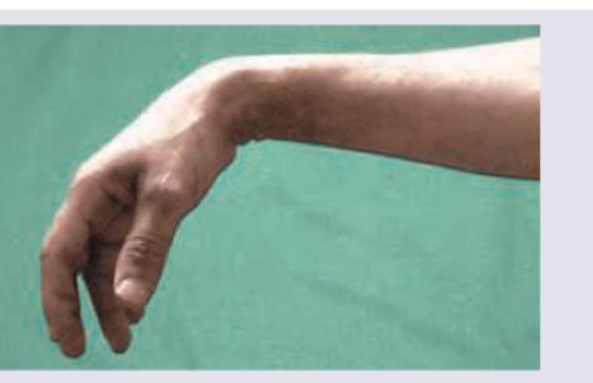
The image shows a clinical finding in a patient's upper limb. Based on these findings, at which anatomical level has the patient most likely sustained a lesion?
Cubital tunnel syndrome occurs due to compression of which structure?
The lower lateral cutaneous nerve of the arm is a branch of which nerve?
Struther's ligament is another name for which of the following?
A cut injury of the ulnar nerve results in what clinical presentation?
Which of the following statements regarding peripheral nerve injuries in the upper limb are true? 1. Radial nerve injury causes anesthesia over the anatomical snuff box. 2. Median nerve injury causes wrist drop. 3. Ulnar nerve injury causes a claw hand. 4. Index finger anesthesia is caused by median nerve injury. 5. Thumb anesthesia is caused by ulnar nerve injury.
Which bursa communicates with the shoulder joint space?
Which of the following two muscles act together for climbing on a tree?
Practice by Chapter
Pectoral Region and Axilla
Practice Questions
Arm and Cubital Fossa
Practice Questions
Forearm and Hand
Practice Questions
Joints of Upper Limb
Practice Questions
Nerves of Upper Limb
Practice Questions
Arterial Supply and Venous Drainage
Practice Questions
Lymphatic Drainage
Practice Questions
Muscles and Their Actions
Practice Questions
Applied Anatomy and Clinical Correlations
Practice Questions
Surface Anatomy and Landmarks
Practice Questions
Want unlimited practice?
Get full access to all questions, explanations, and performance tracking.
Scan to download app