
Upper Limb — MCQs

On this page
A 22-year-old pregnant woman was admitted emergently to the hospital after the baby had begun to appear at the introitus. The baby had presented in the breech position, and it had been necessary to exert considerable traction to complete the delivery. Which of the following structures was most likely injured by the trauma of childbirth?
Clinical testing of the function of the long thoracic nerve is done by:
Paralysis of the opponens muscle leads to the loss of which of the following functions of the thumb?
Abduction of the shoulder is caused by all muscles except:
Which is the main muscle responsible for the opposition of the thumb?
What dermatome provides cutaneous innervation over the medial aspect of the elbow?
A 54-year-old woman is found unconscious. During physical examination, she has an absent biceps brachii reflex. What is the spinal level of the afferent component of this reflex?
Which of the following nerve-muscle/region combinations is FALSE?
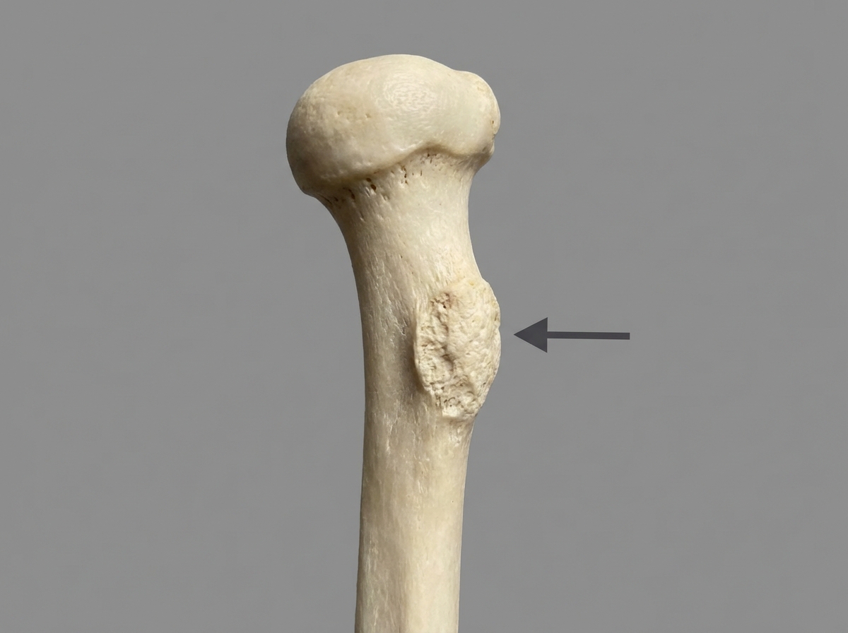
Which muscle is inserted on the area marked by the arrow?
Which of the following is NOT a feature of Quadrangular space syndrome?
Practice by Chapter
Pectoral Region and Axilla
Practice Questions
Arm and Cubital Fossa
Practice Questions
Forearm and Hand
Practice Questions
Joints of Upper Limb
Practice Questions
Nerves of Upper Limb
Practice Questions
Arterial Supply and Venous Drainage
Practice Questions
Lymphatic Drainage
Practice Questions
Muscles and Their Actions
Practice Questions
Applied Anatomy and Clinical Correlations
Practice Questions
Surface Anatomy and Landmarks
Practice Questions
Want unlimited practice?
Get full access to all questions, explanations, and performance tracking.
Scan to download app