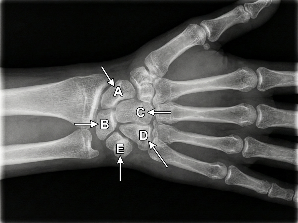
An X-ray of the wrist and hand is shown below. If there is damage to structure E, which of the following muscle weaknesses does it cause?

Which nerve is involved in the Arcade of Frohse?
What is the nerve supply of the trapezius muscle?
What is the nerve supply of the opponens pollicis muscle?
Which of the following statements about the scapula is false?
Pointing index in a supracondylar fracture is due to which nerve injury?
Which muscles produce flexion of the metacarpophalangeal joint?
Which muscle of the hand is NOT supplied by the median nerve?
Following anterior dislocation of the shoulder, a patient develops weakness of flexion at the elbow and lack of sensation over the lateral aspect of the forearm. Which nerve is injured?
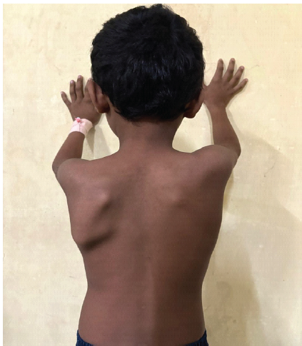
Which ligament prevents the displacement of the humerus?
Explanation: ***Flexor carpi ulnaris*** - The **pisiform bone** (structure E) serves as the primary insertion point for the **flexor carpi ulnaris (FCU)**, which is innervated by the **ulnar nerve**. - Damage to the pisiform disrupts FCU function, causing weakness in **wrist flexion** and **ulnar deviation**, as FCU also inserts on the **hook of hamate** and **5th metacarpal base**. *Flexor carpi radialis* - Innervated by the **median nerve** and inserts on the base of the **2nd metacarpal**, not the pisiform bone. - Pisiform damage would not affect FCR function as it has no anatomical relationship with structure E. *Palmaris longus* - Innervated by the **median nerve** and inserts into the **palmar aponeurosis**, completely bypassing the pisiform bone. - This muscle's function remains intact with pisiform damage as it has no connection to structure E. *Brachioradialis* - Innervated by the **radial nerve** and inserts on the **distal radius**, not involving any carpal bones. - Pisiform bone damage has no impact on brachioradialis function due to its separate anatomical pathway and innervation.
Explanation: The **Arcade of Frohse**, also known as the supinator arch, is the most superior part of the superficial layer of the **supinator muscle**. It is a fibrous, semicircular arch that serves as a critical anatomical landmark in the forearm. **Why the Correct Answer is Right:** The **Posterior Interosseous Nerve (PIN)**, which is the deep terminal branch of the radial nerve, enters the posterior compartment of the forearm by passing beneath this fibrous arch. As the PIN passes through this narrow space between the two heads of the supinator muscle, it is a frequent site of nerve entrapment, leading to **Posterior Interosseous Nerve Syndrome** (characterized by motor weakness of finger and thumb extensors without sensory loss). **Why Other Options are Incorrect:** * **Median Nerve:** This nerve typically passes between the two heads of the **pronator teres** muscle [3] (a common site for Pronator Syndrome) and under the sublimis bridge of the flexor digitorum superficialis [1]. * **Ulnar Nerve:** This nerve passes through the **Cubital Tunnel** (formed by the Osborne’s ligament) behind the medial epicondyle and between the two heads of the flexor carpi ulnaris [1]. * **Radial Nerve:** While the PIN is a branch of the radial nerve, the main trunk of the radial nerve divides into the superficial and deep branches *before* reaching the arcade. The arcade specifically involves the PIN. **High-Yield Clinical Pearls for NEET-PG:** * **PIN Syndrome vs. Radial Nerve Palsy:** PIN syndrome presents with **"Wrist Drop" (incomplete)** or finger drop but **spares sensation**, as the superficial radial nerve (sensory) branches off before the arcade. * The Arcade of Frohse is the most common site for **Radial Tunnel Syndrome**. * **Muscle Innervation:** The PIN innervates all muscles in the posterior compartment of the forearm except the Brachioradialis, Extensor Carpi Radialis Longus (ECRL), and Anconeus (supplied by the main radial nerve) [2].
Explanation: **Explanation:** The **Trapezius** is a large, superficial muscle of the back and neck. It has a unique dual nerve supply that is a frequent high-yield topic in anatomy: 1. **Motor Supply:** The **Spinal Accessory Nerve (CN XI)** provides the motor fibers responsible for muscle contraction. 2. **Sensory Supply:** Branches from the **C3 and C4 spinal nerves** provide proprioceptive fibers. **Analysis of Options:** * **Option D (Correct):** The Spinal Accessory Nerve enters the posterior triangle of the neck and passes deep to the trapezius to supply it. Damage to this nerve results in "drooping of the shoulder" and an inability to shrug. * **Option A (Incorrect):** The **Axillary nerve** (C5-C6) supplies the deltoid and teres minor muscles. * **Option B (Incorrect):** The **Musculocutaneous nerve** (C5-C7) supplies the anterior compartment of the arm (Biceps brachii, Coracobrachialis, and Brachialis). * **Option C (Incorrect):** The **Median nerve** (C5-T1) supplies most of the flexor muscles of the forearm and the thenar muscles of the hand. **High-Yield Clinical Pearls for NEET-PG:** * **Testing:** The trapezius is tested by asking the patient to **shrug their shoulders** against resistance. * **Iatrogenic Injury:** The Spinal Accessory nerve is the most commonly injured nerve during **lymph node biopsies** in the posterior triangle of the neck. * **Action:** It is the primary muscle for **overhead abduction** (above 90 degrees) by rotating the scapula, acting alongside the Serratus Anterior. * **Origin:** It is a "branchiomeric" muscle, meaning it is derived from the pharyngeal arches (specifically the 6th arch), which explains its cranial nerve supply.
Explanation: **Explanation:** The **opponens pollicis** is one of the three muscles forming the thenar eminence of the hand. The correct answer is the **Median nerve** [1], specifically its **recurrent branch** (C8, T1). This nerve supplies the "LOAF" muscles: the lateral two Lumbricals, Opponens pollicis, Abductor pollicis brevis, and Flexor pollicis brevis [3]. **Analysis of Options:** * **Median Nerve (Correct):** It provides motor innervation to the thenar muscles. The recurrent branch is often called the "million-dollar nerve" because its injury results in the loss of thumb opposition, severely disabling hand function [1]. * **Deep branch of the ulnar nerve:** This nerve supplies most of the intrinsic muscles of the hand, including the hypothenar muscles, all interossei, and the Adductor pollicis [3]. It does *not* supply the opponens pollicis. * **Superficial branch of the ulnar nerve:** This is primarily sensory to the medial 1.5 fingers and provides motor supply only to the Palmaris brevis [3]. * **Posterior interosseous nerve:** This is a branch of the radial nerve that supplies the extensor compartment of the forearm; it does not innervate any intrinsic hand muscles [2]. **High-Yield Clinical Pearls for NEET-PG:** * **Ape Thumb Deformity:** Caused by a proximal median nerve injury, leading to atrophy of the thenar eminence and loss of opposition. * **Opponens Digiti Minimi:** Unlike the opponens pollicis, this muscle is supplied by the **deep branch of the ulnar nerve** [3]. * **Mnemonic:** Remember **"Meat LOAF"**—**Me**dian nerve supplies **L**umbricals (1&2), **O**pponens pollicis, **A**bductor pollicis brevis, and **F**lexor pollicis brevis.
Explanation: This question tests your knowledge of the surface anatomy and osteology of the scapula, which is a high-yield topic for NEET-PG. ### **Explanation of the Correct Answer (Option C)** The statement in Option C is **false** because the **medial border** of the scapula does not correspond to the horizontal fissure. Instead, the **oblique fissure** of the lung is indicated by a line drawn from the spine of the scapula (T3) to the 6th costochondral junction. The **horizontal fissure** (present only in the right lung) is represented by a line extending horizontally from the 4th costal cartilage to meet the oblique fissure in the mid-axillary line. ### **Analysis of Other Options** * **Option A:** This is **true**. The root of the spine of the scapula is a standard anatomical landmark located at the level of the **T3 spinous process**. * **Option B:** This is **true**. In the anatomical position, the inferior angle of the scapula typically lies at the level of the **T7 spinous process** (or the 7th intercostal space). * **Option D:** This is **true**. The lateral angle is the thickest part of the scapula and bears the **glenoid cavity**, which articulates with the head of the humerus to form the glenohumeral joint. ### **High-Yield Clinical Pearls for NEET-PG** * **Winging of Scapula:** Caused by injury to the **Long Thoracic Nerve** (Nerve of Bell), leading to paralysis of the **Serratus Anterior**. The medial border becomes prominent. * **Ossification:** The scapula develops from **one primary center** (body) and **seven secondary centers**. The coracoid process has two secondary centers (subcoracoid and coracoid). * **Safe Zone:** The suprascapular nerve passes through the suprascapular notch, a common site for nerve entrapment.
Explanation: **Explanation:** The **pointing index** (also known as the **Kiloh-Nevin sign**) is a classic clinical finding in **Anterior Interosseous Nerve (AIN)** palsy. The AIN is a purely motor branch of the Median nerve. In a supracondylar fracture of the humerus (the most common pediatric elbow fracture), the AIN is the most frequently injured nerve due to its deep position and proximity to the displaced bone fragments. The AIN supplies three muscles: **Flexor Pollicis Longus (FPL)**, the radial half of **Flexor Digitorum Profundus (FDP)** (to the index finger), and Pronator Quadratus [1]. Paralysis of the FPL and FDP prevents flexion of the distal interphalangeal (DIP) joint of the index finger and the interphalangeal (IP) joint of the thumb [1]. When asked to make an "O" sign or a fist, the patient cannot flex these joints, resulting in a "pointing" index finger and a "flat" pinch. **Analysis of Incorrect Options:** * **Radial Nerve:** Injury typically leads to **wrist drop** and inability to extend the fingers/thumb. While it can be injured in supracondylar fractures (especially lateral displacement), it does not cause pointing index. * **Ulnar Nerve:** Injury results in **claw hand** (hyperextension at MCP joints and flexion at IP joints of the ring and little fingers) and weakness of intrinsic hand muscles. * **Musculocutaneous Nerve:** Supplies the coracobrachialis, biceps brachii, and brachialis. Injury leads to weak elbow flexion and loss of sensation on the lateral forearm. **Clinical Pearls for NEET-PG:** * **Most common nerve injured in Supracondylar Fracture:** Anterior Interosseous Nerve (AIN). * **Most common nerve injured in Posteromedial displacement:** Radial Nerve. * **Most common nerve injured in Posterolateral displacement:** Median Nerve. * **AIN Test:** Ask the patient to make the **"OK" sign**. If they cannot form a circle and instead produce a "pulp-to-pulp" pinch, the test is positive for AIN palsy [1].
Explanation: ### Explanation The primary movement of the **Metacarpophalangeal (MCP) joints** involves a coordinated effort between the intrinsic muscles of the hand. The correct answer is **All of the above** because the Lumbricals, Dorsal Interossei, and Palmar Interossei all share a common insertion point that facilitates this specific action [1]. **1. Why the correct answer is right:** The **Lumbricals** and both sets of **Interossei** (Dorsal and Palmar) pass anterior to the transverse axis of the MCP joints before inserting into the **extensor expansions** (dorsal digital expansions) and the bases of the proximal phalanges [1]. Because their tendons cross the MCP joint on the palmar side, their contraction pulls the proximal phalanx into **flexion**. Simultaneously, because they insert into the extensor expansion, they pull the expansion distally, resulting in **extension of the Interphalangeal (IP) joints** [1]. **2. Analysis of Options:** * **Lumbricals:** Originating from the tendons of Flexor Digitorum Profundus, they are the primary muscles for the "Z-movement" (MCP flexion + IP extension) [1]. * **Dorsal Interossei (DAB):** Their primary role is Abduction, but they also contribute significantly to MCP flexion [1]. * **Palmar Interossei (PAD):** Their primary role is Adduction, but like the dorsal set, their anatomical position allows them to assist in MCP flexion [1]. **3. Clinical Pearls & High-Yield Facts for NEET-PG:** * **The "Writing Position":** The combined action of these muscles (Flexion at MCP + Extension at IP) is known as the intrinsic-plus position, essential for holding a pen. * **Ulnar Claw Hand:** Paralysis of these intrinsic muscles (mainly ulnar nerve injury) leads to the opposite deformity: hyperextension at the MCP and flexion at the IP joints. * **Nerve Supply:** All Interossei are supplied by the **Deep branch of the Ulnar nerve**. Lumbricals have a dual supply: 1st and 2nd by the Median nerve, 3rd and 4th by the Ulnar nerve [1].
Explanation: ### Explanation The intrinsic muscles of the hand are primarily supplied by the **Ulnar nerve**, with the exception of five muscles supplied by the **Median nerve** (specifically its recurrent branch and digital branches). These five are remembered by the mnemonic **LOAF**: **L**umbricals (1st and 2nd), **O**pponens pollicis, **A**bductor pollicis brevis, and **F**lexor pollicis brevis. [3] **Why the Question/Options are unique:** There appears to be a technical error in the provided key. **Opponens pollicis (A)**, **Abductor pollicis brevis (B)**, and the **First lumbrical (D)** are all classic examples of muscles **supplied** by the Median nerve. The correct answer to "Which is NOT supplied by the median nerve?" is **C. Extensor pollicis brevis**. This is because all **Extensor** muscles of the thumb and wrist are located in the posterior compartment of the forearm and are supplied by the **Posterior Interosseous Nerve (a branch of the Radial nerve)**. [2] **Analysis of Options:** * **Opponens pollicis & Abductor pollicis brevis:** These are Thenar muscles supplied by the recurrent branch of the median nerve (C8, T1). * **First lumbrical:** Supplied by the digital branches of the median nerve. * **Extensor pollicis brevis:** Supplied by the Radial nerve; it is an extrinsic muscle of the hand. [2] **High-Yield Clinical Pearls for NEET-PG:** * **Ape Thumb Deformity:** Caused by Median nerve injury at the wrist (e.g., Carpal Tunnel Syndrome), leading to paralysis of the thenar muscles. [1] * **Million Dollar Nerve:** The recurrent branch of the median nerve is so named because its injury during carpal tunnel surgery leads to loss of thumb opposition, a major disability. * **Ulnar Paradox:** All interossei and the medial two lumbricals are ulnar-supplied. If the median nerve is spared, the index and middle fingers will not show "clawing."
Explanation: ### Explanation The correct answer is **Musculocutaneous nerve**. **1. Why it is correct:** The musculocutaneous nerve (C5–C7) arises from the lateral cord of the brachial plexus. It is responsible for: * **Motor supply:** It innervates the muscles of the anterior compartment of the arm—**Coracobrachialis, Biceps brachii, and Brachialis**. Loss of these muscles leads to significant weakness in **elbow flexion** and forearm supination. * **Sensory supply:** After passing through the arm, it continues as the **Lateral cutaneous nerve of the forearm**, providing sensation to the lateral aspect of the forearm. In anterior shoulder dislocations, the humeral head can compress or stretch the cords of the brachial plexus, with the musculocutaneous nerve being a classic, though less common, target than the axillary nerve. **2. Why the other options are incorrect:** * **Radial nerve:** Injury (e.g., mid-shaft humerus fracture) leads to "wrist drop" and loss of extension at the elbow/wrist, not flexion weakness. * **Axillary nerve:** This is the *most common* nerve injured in shoulder dislocations. However, it results in deltoid paralysis (loss of abduction) and sensory loss over the "regimental badge" area (lateral shoulder), not the forearm. * **Ulnar nerve:** Injury causes "claw hand" and sensory loss over the medial 1.5 fingers; it does not affect elbow flexion. **3. NEET-PG High-Yield Pearls:** * **Mnemonic for Musculocutaneous muscles:** **BBC** (Biceps, Brachialis, Coracobrachialis). * **Piercing Nerve:** The musculocutaneous nerve is unique because it **pierces the Coracobrachialis muscle**. * **Dislocation Association:** While the **Axillary nerve** is the most frequently injured in shoulder dislocations, the **Musculocutaneous nerve** is the correct clinical fit when forearm sensory loss and flexion weakness are specified.
Explanation: **Explanation:** The **Glenohumeral ligaments** (Superior, Middle, and Inferior) are the primary static stabilizers of the shoulder joint. They are thickenings of the anterior joint capsule that reinforce the joint and prevent the **anterior and inferior displacement** of the humeral head. Among these, the **Inferior Glenohumeral Ligament (IGHL)** is the most important stabilizer when the arm is abducted and externally rotated, acting as a "hammock" to prevent dislocation. **Analysis of Incorrect Options:** * **A. Coracoclavicular:** This ligament (composed of the conoid and trapezoid parts) connects the coracoid process to the clavicle. Its primary function is to stabilize the **acromioclavicular joint** and transmit the weight of the upper limb to the clavicle; it does not directly stabilize the humerus. * **B. Coracohumeral:** While this ligament strengthens the upper part of the capsule, its primary role is to prevent **inferior displacement** of the humerus specifically when the arm is hanging at the side (adducted). It is secondary to the glenohumeral ligaments in overall stability. * **C. Coracoacromial:** This ligament connects two parts of the same bone (scapula). Along with the acromion and coracoid process, it forms the **coracoacromial arch**, which prevents **superior displacement** of the humerus but is not a capsular stabilizer. **NEET-PG High-Yield Pearls:** * The **Inferior Glenohumeral Ligament** is the most frequently injured structure in anterior shoulder dislocations (Bankart lesion). * The shoulder joint is the most commonly dislocated joint in the body due to the disproportionate size between the large humeral head and the shallow glenoid cavity (often compared to a "golf ball on a tee"). * The **Rotator Cuff muscles** (SITS) provide dynamic stability, while the **Glenoid Labrum** deepens the socket to increase static stability.
Pectoral Region and Axilla
Practice Questions
Arm and Cubital Fossa
Practice Questions
Forearm and Hand
Practice Questions
Joints of Upper Limb
Practice Questions
Nerves of Upper Limb
Practice Questions
Arterial Supply and Venous Drainage
Practice Questions
Lymphatic Drainage
Practice Questions
Muscles and Their Actions
Practice Questions
Applied Anatomy and Clinical Correlations
Practice Questions
Surface Anatomy and Landmarks
Practice Questions
Get full access to all questions, explanations, and performance tracking.
Start For Free