
Upper Limb — MCQs

On this page
Which of the following muscles does not have a dual nerve supply?
All of the following muscles are innervated by branches from the brachial plexus except?
Erb's point is formed by which cervical nerve roots?
Which artery lies in Guyon's canal?
What is the lymphatic drainage of the upper outer quadrant of the breast?
Which muscle in the extensor compartment of the forearm causes flexion of the elbow?
What is the nerve supply of the Teres major muscle?
Which muscle crosses the shoulder joint?
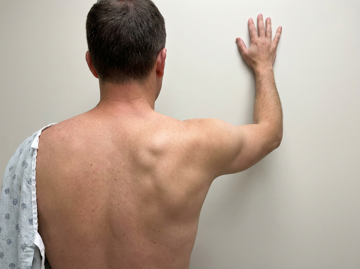
Weakness of which muscle would result in the following type of lesion?
Which of the following muscles is responsible for shrugging the shoulders?
Practice by Chapter
Pectoral Region and Axilla
Practice Questions
Arm and Cubital Fossa
Practice Questions
Forearm and Hand
Practice Questions
Joints of Upper Limb
Practice Questions
Nerves of Upper Limb
Practice Questions
Arterial Supply and Venous Drainage
Practice Questions
Lymphatic Drainage
Practice Questions
Muscles and Their Actions
Practice Questions
Applied Anatomy and Clinical Correlations
Practice Questions
Surface Anatomy and Landmarks
Practice Questions
Want unlimited practice?
Get full access to all questions, explanations, and performance tracking.
Scan to download app