
Upper Limb — MCQs

On this page
Lesions of the musculocutaneous nerve can result in all of the following, EXCEPT:
Which structure is present superficial to the galea aponeurotica (epicranial aponeurosis)?
What is the major blood supply of the pectoralis major muscle?
Damage to the coracobrachialis muscle and to the nerve passing through it could reasonably be expected to produce all of the following, EXCEPT?
Neglected whitlow may lead to necrosis of which part of the distal phalanx?
In a subclavian artery block at the outer border of the 1st rib, all of the following arteries help in maintaining circulation to the upper limb except?
A 22-year-old male football player suffered a wrist injury while falling with force on his outstretched hand. When the anatomic snuffbox is exposed in surgery, an artery is visualized crossing the fractured bone that provides a floor for this space. What artery was visualized?
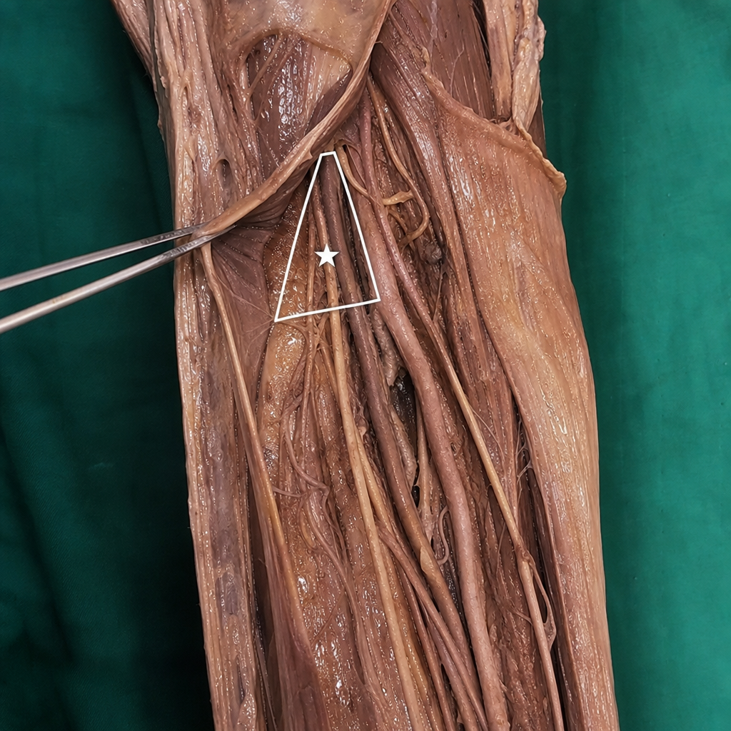
Content of the star marked triangle?
Which of the following statements about the brachial plexus is FALSE?
Guyon's canal is associated with which of the following structures?
Practice by Chapter
Pectoral Region and Axilla
Practice Questions
Arm and Cubital Fossa
Practice Questions
Forearm and Hand
Practice Questions
Joints of Upper Limb
Practice Questions
Nerves of Upper Limb
Practice Questions
Arterial Supply and Venous Drainage
Practice Questions
Lymphatic Drainage
Practice Questions
Muscles and Their Actions
Practice Questions
Applied Anatomy and Clinical Correlations
Practice Questions
Surface Anatomy and Landmarks
Practice Questions
Want unlimited practice?
Get full access to all questions, explanations, and performance tracking.
Scan to download app