
Upper Limb — MCQs

On this page
The circumflex scapular artery is a branch of which structure?
A 23-year-old male soldier accidentally punctured the ventral side of the fifth digit at the base of the distal phalanx while sharpening his knife. The wound became infected and spread into the palm, within the sheath of the flexor digitorum profundus tendons. If the infection were left untreated, into which of the following spaces could it most likely spread?
Erb's palsy occurs due to involvement of which part of the brachial plexus?
Which muscle initiates the abduction of the shoulder?
A physician asks a patient to hold her right upper arm close to her lateral chest wall, and bend the arm at the elbow so that the palm is facing upward. The physician then directs the patient to turn her hand so that the palm faces downward, without bending her wrist. This maneuver causes discomfort to the patient, which the physician notes as pain on?
A 27-year-old male painter presents with inability to abduct his arm more than 15 degrees and difficulty with lateral rotation after a fall. Radiography shows an oblique fracture of the humerus. He also has sensory loss over the shoulder area. Which of the following injuries most likely corresponds to these findings?
Klumpke's paralysis presents with all of the following clinical features except?
A 29-year-old man presents with a stab wound, inability to raise his arm above the horizontal, and a "winged scapula." Which of the following structures of the brachial plexus would most likely be damaged?
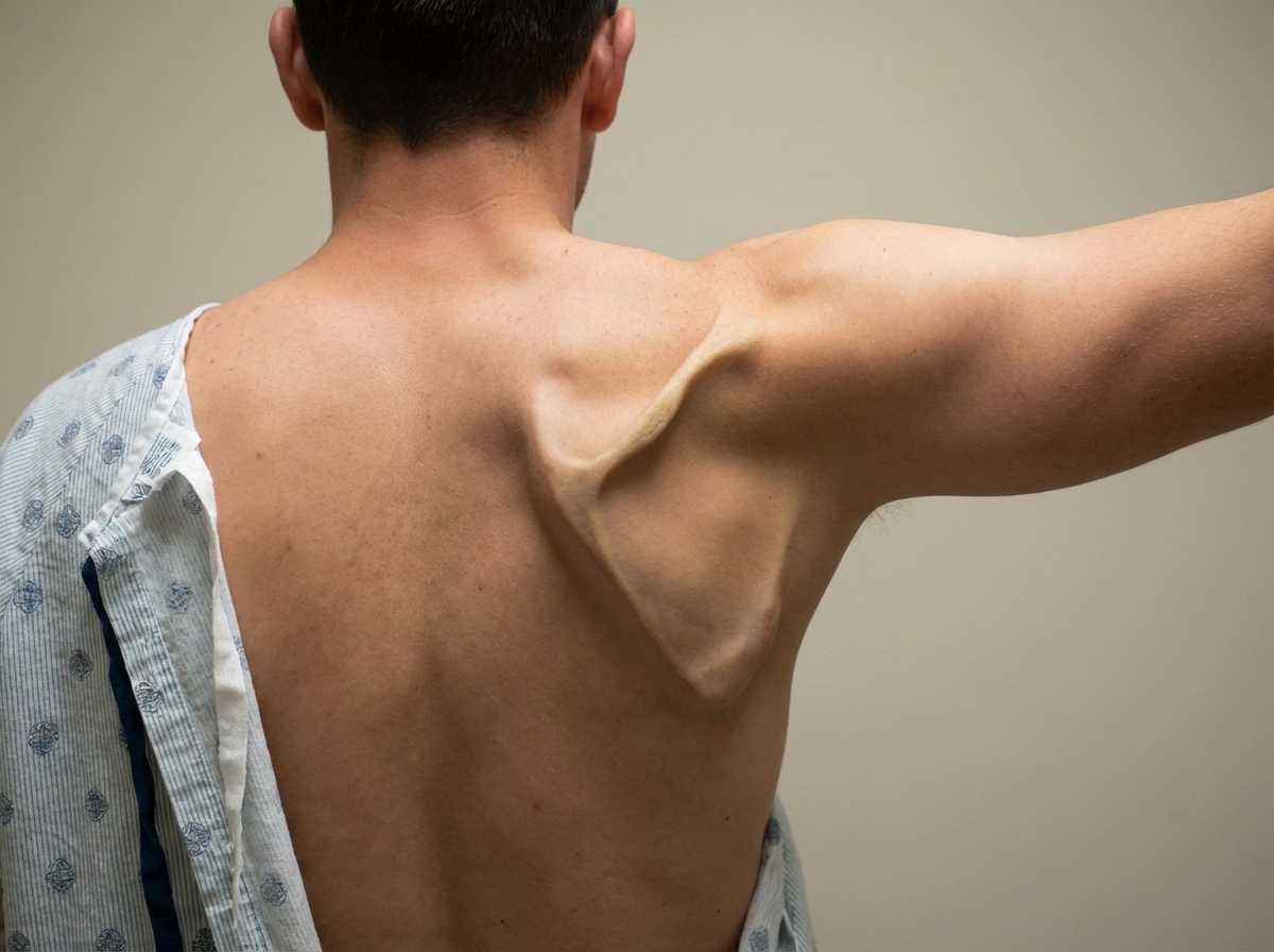
Which nerve is involved in this deformity?
Which spinal nerve roots supply the dermatome for the thumb and index finger region?
Practice by Chapter
Pectoral Region and Axilla
Practice Questions
Arm and Cubital Fossa
Practice Questions
Forearm and Hand
Practice Questions
Joints of Upper Limb
Practice Questions
Nerves of Upper Limb
Practice Questions
Arterial Supply and Venous Drainage
Practice Questions
Lymphatic Drainage
Practice Questions
Muscles and Their Actions
Practice Questions
Applied Anatomy and Clinical Correlations
Practice Questions
Surface Anatomy and Landmarks
Practice Questions
Want unlimited practice?
Get full access to all questions, explanations, and performance tracking.
Scan to download app