
Surface and Radiological Anatomy — MCQs

On this page
On an X-ray chest PA view, which chamber forms the anterior surface of the heart?
The highest point of the iliac crest is at which vertebral level?
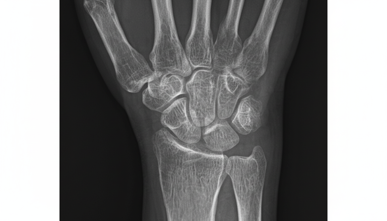
An X-ray of the lower third of the forearm shows which of the following findings?
A thin radiolucent line which follows the root outline on X-rays is?
The mental foramen, which appears as a radiolucency and can be mistaken for periapical pathology, lies close to the apex of which tooth?
In a standing man, at the midaxillary line, where does the lower border of the pleura reach?
Which of the following is TRUE regarding the level of the superficial palmar arch?
Radiographically, the mental foramen lies between the roots of which teeth?
What anatomical landmark is described by Shenton's line?
A 33-year-old male is admitted to the hospital after a violent, multiple car collision. His blood pressure is 89/39 mm Hg, and a central venous line is ordered to be placed. Which of the following structures is used as a landmark to place the tip of the catheter of the central venous line?
Practice by Chapter
Surface Landmarks of the Head and Neck
Practice Questions
Surface Landmarks of the Thorax
Practice Questions
Surface Landmarks of the Abdomen
Practice Questions
Surface Landmarks of the Limbs
Practice Questions
Radiographic Anatomy
Practice Questions
CT Anatomy
Practice Questions
MRI Anatomy
Practice Questions
Ultrasonographic Anatomy
Practice Questions
Angiographic Anatomy
Practice Questions
Sectional and Cross-sectional Anatomy
Practice Questions
Anatomical Correlations in Common Imaging
Practice Questions
Interventional Radiological Anatomy
Practice Questions
Want unlimited practice?
Get full access to all questions, explanations, and performance tracking.
Scan to download app