What is the normal anteroposterior length of the eyeball?
The inferior orbital fissure is located between which two walls of the orbit?
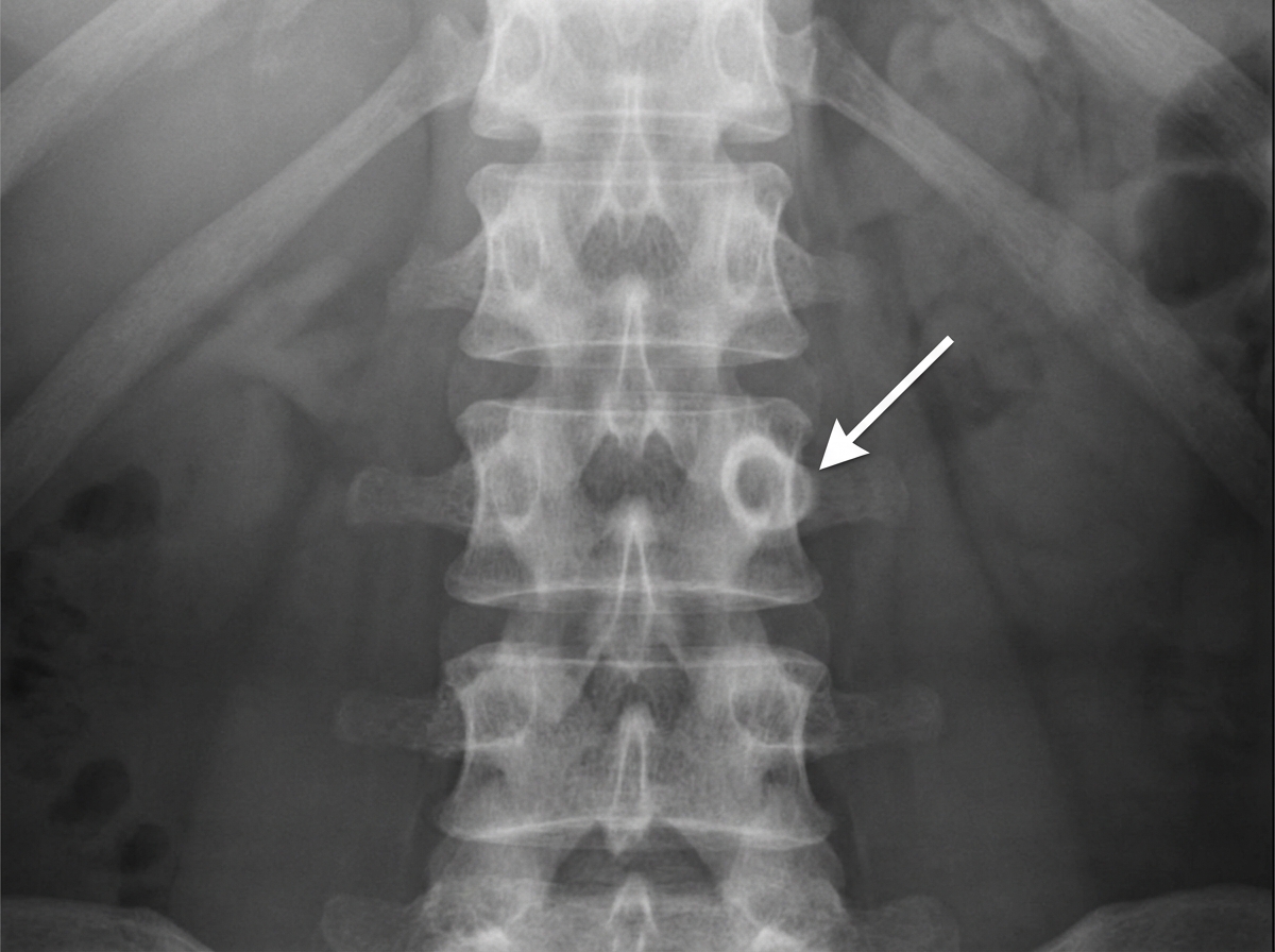
Which anatomical structure is characterized by a worm-hole radiolucency?
Sappey's line denotes a line encircling which anatomical region?
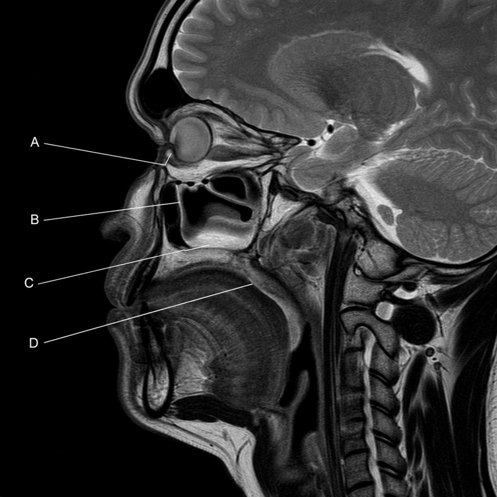
In an MRI scan showing a sagittal section through the head and neck, tears drain through the nasolacrimal duct into the space below which structure?
What is the term for the area of skin supplied by a single nerve?
What is true regarding the surface anatomy of the internal jugular vein?
Which of the following anatomical structures is NOT typically found at the transpyloric plane?
The umbilicus is supplied by which spinal segment?
Surface marking of the internal jugular vein is obtained by joining which two points?
Explanation: The eyeball is an asymmetrical sphere housed within the bony orbit. Its dimensions are critical in clinical ophthalmology, particularly for calculating intraocular lens power and diagnosing refractive errors. **1. Why 24 mm is correct:** The **anteroposterior (axial) diameter** of a normal adult human eyeball is approximately **24.2 mm** (commonly rounded to **24 mm**). This measurement represents the distance from the anterior pole (cornea) to the posterior pole (sclera) [1]. * **Vertical diameter:** ~23 mm * **Transverse diameter:** ~23.5 mm The eyeball is slightly shorter vertically than it is wide or long, making it an "oblate spheroid." **2. Analysis of incorrect options:** * **12 mm (A):** This is roughly the diameter of the **cornea** (horizontal diameter is ~11.7 mm). * **16 mm (B):** This is the approximate axial length of a **newborn's eyeball**. It grows rapidly in the first two years of life. * **20 mm (C):** An axial length this short in an adult would result in severe **Hypermetropia** (farsightedness), as the image focuses behind the retina [1]. **3. Clinical Pearls for NEET-PG:** * **Refractive Errors:** A 1 mm increase in axial length (longer than 24 mm) results in approximately **-3 Diopters of Myopia** (nearsightedness) [1]. Conversely, a shorter eyeball leads to Hypermetropia. * **Volume:** The total volume of the adult eyeball is approximately **6.5 mL**. * **Weight:** The eyeball weighs approximately **7 grams**. * **Coat Thickness:** The sclera is thickest posteriorly (1 mm) and thinnest at the insertion of extraocular muscles (0.3 mm).
Explanation: The orbit is a pyramidal cavity formed by seven bones. Understanding the junctions between its four walls is crucial for identifying key neurovascular passages. ### **Explanation of the Correct Answer** The **Inferior Orbital Fissure (IOF)** is located at the junction of the **lateral wall and the floor** of the orbit. * **Boundaries:** It is bounded superiorly by the greater wing of the sphenoid (lateral wall) and inferiorly by the maxilla and orbital process of the palatine bone (floor). * **Function:** It connects the orbit to the pterygopalatine and infratemporal fossae, transmitting the maxillary nerve (V2), zygomatic nerve, infraorbital vessels, and the inferior ophthalmic vein. ### **Analysis of Incorrect Options** * **A & D (Roof and Medial/Lateral Wall):** The roof is primarily formed by the frontal bone. The junction between the roof and the lateral wall contains the **Superior Orbital Fissure (SOF)**, not the inferior. * **C (Floor and Medial Wall):** This junction is relatively continuous, formed by the maxilla and ethmoid bones. The most significant structure in this vicinity is the **nasolacrimal canal**, located anteriorly. ### **High-Yield Clinical Pearls for NEET-PG** * **Superior Orbital Fissure (SOF):** Located between the **roof (lesser wing of sphenoid)** and **lateral wall (greater wing)**. It transmits CN III, IV, V1 (lacrimal, frontal, nasociliary), and VI. * **Blow-out Fracture:** Most commonly involves the **floor** (weakest point). If the fracture extends to the IOF, it can cause anesthesia in the distribution of the infraorbital nerve. * **Optic Canal:** Located in the **lesser wing of the sphenoid** (superomedial aspect of the orbital apex), transmitting the Optic nerve and Ophthalmic artery.
Explanation: ### Explanation **Correct Answer: C. Nutrient canal** **Anatomical Concept:** Nutrient canals are small, tunnel-like passages in the alveolar bone that house blood vessels and nerves supplying the teeth and gingiva. On a radiograph (especially intraoral periapical views), they appear as thin, vertical, linear radiolucencies. When these canals are viewed in a specific orientation or are particularly prominent in the mandibular anterior region (where the bone is thin), they present as small, dark, circular spots or linear streaks. This characteristic radiographic appearance is classically described as **"worm-hole" radiolucency.** **Analysis of Incorrect Options:** * **A. Maxillary sinus:** This appears as a large, well-defined, radiolucent "pneumatized" cavity in the maxilla, often bordered by a thin white line (cortical border). It does not exhibit a "worm-hole" pattern. * **B. Mandibular canal:** This is a significant, continuous radiolucent band bounded by radiopaque cortical lines, housing the inferior alveolar nerve and vessels. It is much larger and more structured than nutrient canals. * **D. Mental foramen:** This typically appears as a single, well-defined, oval or round radiolucency located near the apices of the mandibular premolars. **High-Yield Clinical Pearls for NEET-PG:** * **Location:** Nutrient canals are most commonly seen in the **mandibular incisor region** and the walls of the maxillary sinus. * **Clinical Significance:** They are more prominent in patients with **periodontal disease** or **thin alveolar bone** (senile atrophy). * **Differential Diagnosis:** They should not be confused with fracture lines or periapical pathology. Unlike fractures, nutrient canals have smooth, cortical borders and follow a predictable anatomical path. * **Radiographic Landmark:** They are often seen as vertical lines between the roots of the mandibular incisors.
Explanation: **Sappey’s line** is a critical landmark in lymphatic anatomy, specifically related to the drainage of the skin [1]. It refers to a horizontal line encircling the trunk, passing approximately **2 cm above the umbilicus** (transumbilical line) and extending to the level of the second lumbar vertebra posteriorly [1]. ### Why the correct answer is right: The significance of Sappey’s line lies in the **watershed area** of cutaneous lymphatic drainage. * **Above the line:** Lymph from the skin drains upwards into the **axillary lymph nodes** [1]. * **Below the line:** Lymph from the skin drains downwards into the **superficial inguinal lymph nodes** [1]. The umbilicus itself is a unique site where these drainage patterns meet, making Sappey’s line the functional boundary between the upper and lower trunk lymphatics. ### Why the incorrect options are wrong: * **Option A (Neck at C6):** This level is significant for the cricoid cartilage, the beginning of the trachea/esophagus, and the carotid tubercle (Chassaignac's tubercle), but it has no association with Sappey’s line. * **Option C (Salpingian tubes):** While Marie Philibert Constant Sappey was an anatomist who studied various structures, "Sappey’s line" specifically refers to the trunk's lymphatic watershed, not the Fallopian (salpingian) tubes. ### High-Yield Clinical Pearls for NEET-PG: * **Clinical Application:** Understanding Sappey’s line is vital for predicting the **metastatic spread** of skin cancers (like melanoma) on the trunk. * **Sappey’s Plexus:** Do not confuse the line with the subareolar lymphatic plexus of the breast, also named after Sappey. * **Vertical Watershed:** The midline of the body acts as a vertical watershed; lymph rarely crosses the midline under normal physiological conditions.
Explanation: The nasolacrimal duct (NLD) is the terminal part of the lacrimal apparatus responsible for draining tears from the lacrimal sac into the nasal cavity. Anatomically, it opens into the **inferior meatus** of the nose. * **Correct Option (Structure B):** In a sagittal MRI, Structure B represents the **Inferior Nasal Concha (Turbinate)**. The space located immediately below and lateral to this structure is the **inferior meatus**. The nasolacrimal duct pierces the lateral wall of this meatus to drain tears. This opening is partially guarded by a mucosal fold known as **Hasner’s valve** (lacrimal fold), which prevents air from being blown into the lacrimal sac. * **Incorrect Options:** * **Structure A (Superior Concha):** The space below this is the superior meatus, which receives the posterior ethmoidal air cells. * **Structure C (Middle Concha):** The space below this is the middle meatus, which is a high-yield area containing the hiatus semilunaris. It receives drainage from the frontal, maxillary, and anterior/middle ethmoidal sinuses. * **Structure D (Spheno-ethmoidal recess):** Located above and behind the superior concha, it receives the drainage of the sphenoid sinus. **High-Yield Clinical Pearls for NEET-PG:** * **Epiphora:** Excessive tearing often caused by obstruction of the NLD. * **Dacryocystitis:** Infection of the lacrimal sac, usually secondary to NLD obstruction. * **Development:** The NLD is the last part of the lacrimal system to canalize; failure of canalization at the inferior meatus leads to congenital NLD obstruction in newborns.
Explanation: ### Explanation **Correct Option: A. Dermatome** A **dermatome** is defined as an area of skin supplied by the sensory fibers of a **single spinal nerve** (via its dorsal root). These areas are arranged in a segmental fashion along the body. Understanding dermatomes is crucial for localizing spinal cord injuries or nerve root compressions (radiculopathy). For example, loss of sensation at the level of the umbilicus points to a lesion at the **T10** spinal level. **Incorrect Options:** * **B. Pedicle:** In anatomy, a pedicle is a stalk-like structure that connects an organ or tissue to the body (e.g., the bony pillars of a vertebral arch or the vascular stalk used in reconstructive surgery). * **C. Graft:** This refers to a piece of living tissue (skin, bone, or organ) that is surgically moved from one site to another to replace or repair damaged tissue. Unlike a flap, a graft does not have its own blood supply. * **D. Dermoid:** This usually refers to a **dermoid cyst**, a type of germ cell tumor (teratoma) containing mature skin, hair follicles, and sweat glands. **High-Yield Clinical Pearls for NEET-PG:** * **C6:** Thumb; **C7:** Middle finger; **C8:** Little finger. * **T4:** Nipple line; **T10:** Umbilicus. * **L1:** Inguinal ligament; **L4:** Medial malleolus and big toe. * **S1:** Lateral malleolus and little toe. * **Herpes Zoster (Shingles):** This virus remains latent in the dorsal root ganglion and manifests as a painful vesicular rash strictly following a specific dermatomal distribution. * **Note:** While dermatomes represent a single spinal nerve, a **myotome** refers to the group of muscles supplied by a single spinal nerve.
Explanation: **Explanation:** The **Internal Jugular Vein (IJV)** is the largest vein in the neck, collecting blood from the brain, face, and neck. Understanding its surface projection is critical for clinical procedures like central venous catheterization. **1. Why Option B is Correct:** The surface anatomy of the IJV is represented by a broad line joining two points: * **Upper point:** The lobule of the ear (representing the jugular bulb just below the jugular foramen). * **Lower point:** The medial end of the clavicle (specifically the sternoclavicular joint), where the IJV joins the subclavian vein to form the brachiocephalic vein. The vein lies deep to the sternocleidomastoid (SCM) muscle, specifically within the triangle formed by the sternal and clavicular heads of the SCM. **2. Why Other Options are Incorrect:** * **Options A & D:** The **midpoint of the clavicle** is the surface landmark for the **External Jugular Vein (EJV)**, which crosses the SCM obliquely. * **Option C:** The **lateral end of the clavicle** does not correspond to any major vertical vascular structure in the neck. * **Option D:** While the mastoid process is near the ear lobule, the IJV specifically aligns with the lobule to reach the medial clavicle; the mastoid to midpoint line is an incorrect hybrid of IJV and EJV landmarks. **High-Yield NEET-PG Pearls:** * **Relation to Carotid:** In the carotid sheath, the IJV is **lateral** to the common carotid artery. * **Cannulation:** The IJV is preferred over the subclavian vein for central lines because it has a straighter path to the right atrium and a lower risk of pneumothorax. * **Right vs. Left:** The **Right IJV** is preferred for catheterization as it is larger and forms a direct vertical line with the superior vena cava. **Note:** None of the provided references [1, 2, 3, 4, 5] contain information regarding the surface anatomy landmarks of the Internal Jugular Vein (ear lobule to medial clavicle), therefore no inline citations were added to the explanation text to maintain accuracy.
Explanation: The **Transpyloric Plane (Addison’s Plane)** is a key anatomical landmark located midway between the suprasternal notch and the pubic symphysis (at the level of the **L1 vertebra**). ### **Why "Body of the gallbladder" is the correct answer:** The **Fundus** of the gallbladder is the specific part that lies at the transpyloric plane, specifically where the lateral border of the rectus abdominis muscle (linea semilunaris) meets the 9th costal cartilage. The **Body** of the gallbladder is situated more superiorly and posteriorly against the liver [1], making it an incorrect association for this specific plane. ### **Analysis of Incorrect Options:** * **Pylorus of the stomach:** This is the namesake of the plane. In a supine position, the pylorus lies at the level of L1. * **Hilum of the kidney:** The transpyloric plane passes through the hila of both kidneys—specifically the **upper part of the left hilum** and the **lower part of the right hilum** (due to the liver pushing the right kidney lower). * **Neck of the pancreas:** The transpyloric plane passes directly through the neck of the pancreas. The head lies below it, and the body/tail lie slightly above it. ### **NEET-PG High-Yield Pearls:** To master questions on the Transpyloric Plane (L1), remember the mnemonic **"P-H-A-N-T-O-M-S"**: * **P:** **P**ylorus of stomach, **P**ancreas (neck). * **H:** **H**ila of kidneys. * **A:** **A**rtery (Origin of Superior Mesenteric Artery). * **N:** **N**inth costal cartilage. * **T:** **T**ermination of spinal cord (Conus Medullaris). * **O:** **O**ddi (Sphincter of Oddi). * **M:** **M**idpoint between jugular notch and pubic symphysis. * **S:** **S**pleen (upper pole), **S**econd part of duodenum (junction with first). **Clinical Note:** This plane also marks the origin of the portal vein and the cisterna chyli.
Explanation: ### Explanation The sensory innervation of the skin of the anterior abdominal wall is provided by the anterior rami of the lower six thoracic nerves (T7–T12) and the first lumbar nerve (L1). These follow a segmental distribution known as **dermatomes** [2]. **Why T10 is correct:** The **T10 dermatome** specifically encircles the abdomen at the level of the **umbilicus** [3]. This is a classic anatomical landmark used in clinical examinations to localize spinal cord levels or sensory deficits [3]. **Analysis of Incorrect Options:** * **T4:** This dermatome is located at the level of the **nipples** (teat = T4) in males and prepubescent females. * **T6:** This dermatome corresponds to the level of the **xiphoid process** of the sternum [3]. * **T8:** This dermatome supplies the skin of the upper abdomen, roughly halfway between the xiphoid process and the umbilicus. **Clinical Pearls for NEET-PG:** 1. **Referred Pain:** Early appendicitis pain is often felt in the periumbilical region because the appendix and the T10 dermatome share the same spinal segment for visceral and somatic afferents [3]. 2. **L1 Landmark:** The inguinal ligament and the pubic symphysis region are supplied by the **L1** nerve (via iliohypogastric and ilioinguinal nerves) [2]. 3. **Lymphatic Drainage:** The umbilicus acts as a watershed line. Lymph from the skin *above* the umbilicus drains into the **axillary lymph nodes**, while lymph from *below* the umbilicus drains into the **superficial inguinal lymph nodes** [1]. 4. **Caput Medusae:** The umbilicus is a site of **porto-systemic anastomosis** between the paraumbilical veins (portal) and the superficial epigastric veins (systemic).
Explanation: The **Internal Jugular Vein (IJV)** is the largest vein in the neck, collecting blood from the brain, face, and neck. To mark its surface projection, a line is drawn connecting two specific points: 1. **Superior point:** A point on the neck just medial to the **lobule of the ear** (representing the jugular foramen at the base of the skull). 2. **Inferior point:** The **medial end of the clavicle** (specifically the depression between the sternal and clavicular heads of the sternocleidomastoid muscle), which corresponds to the location of the brachiocephalic vein formation. **Analysis of Options:** * **Option A (Correct):** Accurately describes the anatomical course from the skull base to the root of the neck. * **Option B:** This describes the surface marking of the **Common Carotid Artery**, which lies medial to the IJV. The point between the mastoid and the angle of the mandible corresponds to the bifurcation of the carotid. * **Option C:** This does not correspond to any major neurovascular structure; the IJV ends behind the sternoclavicular joint, but it does not extend to the middle of the clavicle. * **Option D:** This is the surface marking for the **External Jugular Vein**, which runs superficially across the sternocleidomastoid muscle from the angle of the mandible to the midpoint of the clavicle. **Clinical Pearls for NEET-PG:** * **Central Venous Pressure (CVP):** The right IJV is preferred for CVP monitoring because it is in a direct vertical line with the Superior Vena Cava and Right Atrium. * **Cannulation:** During IJV cannulation, the needle is typically inserted at the apex of the triangle formed by the two heads of the sternocleidomastoid muscle, directed towards the ipsilateral nipple. * **Relation:** The IJV lies **lateral** to the Common Carotid Artery within the carotid sheath.
Explanation: The **Internal Jugular Vein (IJV)** is a major venous channel in the neck, and its surface marking is a high-yield topic for both anatomy and clinical procedures like central venous pressure (CVP) monitoring. ### **Explanation of the Correct Answer** The surface marking of the IJV is represented by a broad line connecting two points: 1. **Superior Point:** The lobule of the ear (representing the jugular bulb just below the jugular foramen). 2. **Inferior Point:** The medial end of the clavicle (specifically the sternoclavicular joint). The vein descends vertically within the **carotid sheath**, lateral to the internal and common carotid arteries, and deep to the sternocleidomastoid (SCM) muscle. It ends behind the medial end of the clavicle by joining the subclavian vein to form the brachiocephalic vein. ### **Analysis of Incorrect Options** * **Option A & D:** The **midpoint of the clavicle** is the surface marking for the **Subclavian Vein** (where it is accessed for catheterization). * **Option C:** The **lateral end of the clavicle** does not correspond to any major vertical vascular structure in the neck. * **Note on External Jugular Vein (EJV):** The EJV is marked by a line from the angle of the mandible to the midpoint of the clavicle, crossing the SCM obliquely. ### **NEET-PG High-Yield Pearls** * **Relation to SCM:** The IJV lies deep to the SCM. Its lower end is found in the **lesser supraclavicular fossa** (the triangle between the sternal and clavicular heads of the SCM). * **Clinical Use:** The IJV is preferred for central line insertion because it has a straight course to the right atrium and lacks valves (allowing for JVP assessment). * **Right vs. Left:** The Right IJV is usually preferred for cannulation as it is larger and provides a more direct path to the Superior Vena Cava.
Explanation: The esophagus is a muscular tube approximately **25 cm** in length. However, in clinical practice (such as endoscopy), measurements are taken from the **upper incisor teeth**. The gastroesophageal (GE) junction is located at the level of the **T11 vertebra**, which corresponds to a distance of approximately **40 cm** from the incisors. [1] **Breakdown of Distances from Upper Incisors:** * **15 cm:** This marks the **commencement of the esophagus** at the cricopharyngeal sphincter (lower border of the cricoid cartilage/C6 level). This is the narrowest point of the esophagus. On endoscopy, the cervical esophagus corresponds to approximately 15 to 20 cm from the incisors. [1] * **25 cm:** This corresponds to the level where the **arch of the aorta and the left main bronchus** cross the esophagus (T4/T5 level). This is approximately 20 to 25 cm from the incisors. [1] * **40 cm:** This is the distance to the **gastroesophageal junction**, where the esophagus pierces the diaphragm (T10) and enters the stomach (T11). [1] **Why the other options are incorrect:** * **15 cm:** Too proximal; represents the esophageal inlet. [1] * **25 cm:** Represents the mid-esophagus/broncho-aortic constriction. [1] * **60 cm:** Too distal; this distance would typically reach the pylorus or the first part of the duodenum. **Clinical Pearls for NEET-PG:** 1. **Constrictions:** Remember the "15-25-40" rule for the three major anatomical constrictions (Cervical, Thoracic, and Diaphragmatic). Some texts include a fourth at 22 cm (Aortic arch). 2. **Vertebral Levels:** Esophagus starts at **C6**, pierces the diaphragm at **T10**, and ends at **T11**. 3. **Portosystemic Anastomosis:** The GE junction is a critical site for portosystemic shunting; esophageal varices develop here in portal hypertension due to communication between the left gastric vein and the azygos vein.
Explanation: To master surface anatomy for NEET-PG, it is crucial to distinguish between the **anatomical location** (where the valve is) and the **auscultatory area** (where the sound is best heard). [1] ### **1. Why Option A is Correct** The **aortic valve** is anatomically situated behind the left half of the sternum, at the level of the **lower border of the left 3rd costal cartilage** and the adjoining part of the sternum. [1] It is a semilunar valve that prevents backflow from the aorta into the left ventricle. ### **2. Analysis of Incorrect Options** * **Option B (Right 3rd costal cartilage):** This is the anatomical location of the **Superior Vena Cava** entering the right atrium, not the aortic valve. * **Option C (Right 2nd intercostal space):** This is the **Auscultatory Area** for the aortic valve. Sound is conducted here by the column of blood in the ascending aorta, which is closest to the chest wall at this point. * **Option D (Right 3rd intercostal space):** This does not correspond to a primary cardiac valve landmark. ### **3. High-Yield Clinical Pearls for NEET-PG** To remember the anatomical positions of the four valves, use the mnemonic **"P-M-A-T"** (Pulmonary, Mitral, Aortic, Tricuspid) from superior to inferior: * **Pulmonary Valve:** Left 3rd costal cartilage (Upper border). [1] * **Aortic Valve:** Left 3rd costal cartilage (Lower border). [1] * **Mitral Valve:** Left 4th costal cartilage. * **Tricuspid Valve:** Right/Middle of sternum at the level of the 4th/5th intercostal space. **Key Distinction:** If a question asks for the **site of auscultation**, the answer is the Right 2nd Intercostal Space. If it asks for **surface marking/anatomical position**, the answer is the Left 3rd Costal Cartilage.
Explanation: The **hilar shadow** on a standard frontal chest X-ray is a complex opacity formed primarily by the vascular and bronchial structures entering and leaving the lungs. Understanding its composition is high-yield for radiological anatomy [1]. ### Why Lower Lobe Pulmonary Veins is the Correct Answer: The hilar shadow is predominantly formed by the **pulmonary arteries** and the **upper lobe pulmonary veins**. The **lower lobe pulmonary veins** do not contribute to the hilum because they enter the left atrium at a level **inferior and posterior** to the hilum. On a chest radiograph, these veins are seen crossing the lower lung fields toward the heart, separate from the main hilar opacity. ### Analysis of Incorrect Options: * **A. Pulmonary Arteries:** These are the primary contributors to the hilar shadow. The right pulmonary artery and the left pulmonary artery (which arches over the left main bronchus) form the bulk of the density seen on X-ray. * **B. Upper Lobe Pulmonary Veins:** These veins pass through the hilar region to reach the superior aspect of the left atrium, contributing significantly to the superior and lateral margins of the hilar shadow. * **D. Lobar Bronchi:** While air-filled and less dense than vessels, the walls of the major bronchi and the surrounding connective tissue/lymph nodes contribute to the overall structural volume of the hilum [1]. ### High-Yield NEET-PG Pearls: * **The "Left is Higher" Rule:** The left hilum is normally higher than the right hilum in 95% of individuals because the left pulmonary artery arches over the left main bronchus. * **Hilar Point:** This is the angle formed by the intersection of the superior pulmonary vein and the descending pulmonary artery. Displacement of this point is a sensitive sign of lung collapse or masses. * **Vascular Dominance:** Remember that nearly **80% of the hilar density** is vascular (arteries > veins).
Explanation: The **Internal Jugular Vein (IJV)** is a vital vascular structure in the neck, and its surface projection is a high-yield topic for clinical procedures like central venous catheterization. **1. Why Option A is Correct:** The surface marking of the IJV is represented by a line connecting two points: * **Upper point:** The **ear lobule** (which corresponds to the level of the jugular foramen at the skull base). * **Lower point:** The **medial end of the clavicle** (specifically the sternoclavicular joint). The vein descends vertically behind the Sternocleidomastoid (SCM) muscle, lying lateral to the Internal and Common Carotid arteries within the carotid sheath. **2. Analysis of Incorrect Options:** * **Option B & C:** The **middle and lateral thirds of the clavicle** are landmarks for the **External Jugular Vein (EJV)**. The EJV runs obliquely across the SCM muscle and pierces the deep fascia above the middle third of the clavicle to join the subclavian vein. * **Option D:** While the **mastoid process** is near the ear lobule, the standard anatomical landmark for the IJV's superior extent in surface marking is the lobule. The mastoid process is more commonly used as a landmark for the insertion of the SCM muscle. **3. Clinical Pearls for NEET-PG:** * **Cannulation:** The IJV is typically accessed in the "Sedillot’s triangle"—the space between the sternal and clavicular heads of the SCM muscle. * **Right vs. Left:** The **Right IJV** is preferred for central line placement because it is larger, more superficial, and provides a straight path to the Right Atrium. * **JVP:** The Internal Jugular Vein (not the External) is used to measure **Jugular Venous Pressure** because it lacks valves and is in direct continuity with the Right Atrium.
Explanation: The fovea centralis is a small, specialized pit located at the center of the macula lutea, responsible for sharp central vision. In ophthalmoscopy, its position is measured relative to the optic disc (the anatomical "blind spot") [1]. 1. Why Option B is correct: Anatomically, the fovea is located approximately 3.0 mm (or 2 disc diameters) temporal to the temporal margin of the optic disc. It also lies slightly inferior (about 0.5–1.0 mm) to the horizontal meridian of the disc [1]. Since the average diameter of an optic disc is roughly 1.5 mm, a 3.0 mm distance equates precisely to 2 disc diameters (DD). 2. Why other options are incorrect: * Option A (1 DD): This distance is too short; at 1 DD, you would still be within the peripapillary region of the retina. * Options C & D (3 and 4 DD): These distances are too far temporal. At 3–4 disc diameters away, you would be moving toward the peripheral retina, far beyond the macula. High-Yield NEET-PG Clinical Pearls: * The Macula: Measures approximately 5.5 mm in diameter. * The Fovea: Measures approximately 1.5 mm in diameter (the same size as the optic disc). * Foveola: The central-most part of the fovea (0.35 mm); it is the thinnest part of the retina and contains only cones (no rods, no blue cones). * Foveal Avascular Zone (FAZ): Located within the fovea; it is crucial for high-acuity vision and is easily identified on Fundus Fluorescein Angiography (FFA). * Clinical Landmark: In cases of central retinal artery occlusion (CRAO), the "cherry-red spot" appears at the fovea because the underlying choroid shows through the thin retinal layers.
Explanation: **Explanation:** The **optic disc** (optic nerve head) is the anatomical location where ganglion cell axons exit the eye to form the optic nerve [1]. It is a vertically oval structure with an average diameter of approximately **1.5 mm**. Understanding this dimension is crucial for clinical fundoscopy, as the optic disc serves as the standard "unit of measurement" (Disc Diameter) for locating other retinal lesions. **Analysis of Options:** * **Option B (1.5 mm):** This is the correct anatomical average. While it can range from 1.2 mm to 1.8 mm, 1.5 mm is the standard value used in medical examinations. * **Option A (0.5 mm):** This is too small for the disc; however, it is approximately the diameter of the **foveola** (the central-most floor of the fovea). * **Option C (5.5 mm):** This is the approximate diameter of the entire **macula lutea**. * **Option D (10.5 mm):** This value has no anatomical correlation with the posterior pole of the eye; the entire eyeball diameter is only about 24 mm. **High-Yield Clinical Pearls for NEET-PG:** * **Blind Spot:** The optic disc lacks photoreceptors (rods and cones), corresponding to the physiological blind spot in the visual field [1]. * **Location:** It is situated 3–4 mm nasal to the fovea. * **Cup-Disc Ratio:** A normal ratio is < 0.3. An increase in this ratio (cupping) is a hallmark sign of **Glaucoma** [2]. * **Papilledema:** This refers to bilateral optic disc swelling due to increased intracranial pressure. On fundoscopy, it presents with blurring of disc margins and loss of venous pulsations.
Explanation: **Explanation:** The correct answer is **T10**. This question tests the knowledge of dermatomes—the specific areas of skin supplied by a single spinal nerve root. **1. Why T10 is correct:** The umbilicus is a key anatomical landmark used to map the sensory distribution of the thoracic spinal nerves [1]. The **T10 dermatome** consistently supplies the skin at the level of the umbilicus [1]. This is a crucial reference point in clinical examinations to localize spinal cord injuries or determine the level of spinal anesthesia. **2. Analysis of Incorrect Options:** * **T4:** Supplies the skin at the level of the **nipples** (intermammary line). * **T6:** Supplies the skin at the level of the **xiphoid process** [1]. * **T8:** Supplies the upper abdominal wall, midway between the xiphoid process and the umbilicus. **3. Clinical Pearls & High-Yield Facts for NEET-PG:** * **Referred Pain:** In early appendicitis, visceral pain is referred to the **T10 (umbilical) region** because the appendix and the umbilicus share the same spinal segment for sensory fibers [1]. As the parietal peritoneum becomes inflamed, the pain shifts to the Right Iliac Fossa (McBurney’s point) [1]. * **L1 Dermatome:** Supplies the skin over the **inguinal ligament** and the pubic symphysis [1]. * **C3, C4, C5:** "Keep the diaphragm alive" (Phrenic nerve), but the **C4** dermatome also covers the "cape" of the shoulder (supraclavicular nerves). * **Beevor’s Sign:** A clinical test where the umbilicus moves upward when a patient attempts to sit up; it indicates paralysis of the lower abdominal muscles (T10-T12) with sparing of the upper ones, often seen in spinal cord lesions at the T10 level.
Explanation: The movement of abdominal organs during respiration is primarily determined by their relationship to the **diaphragm**. When the diaphragm contracts during inspiration, it descends, pushing the underlying viscera downward. **Why Pancreas is the Correct Answer:** The **Pancreas** is a **retroperitoneal organ** (except for the tail) that is firmly fixed to the posterior abdominal wall. Unlike the liver or stomach, it lacks a mesentery that would allow for significant mobility. More importantly, it is not directly attached to the diaphragm. Its fixed position behind the peritoneum and its attachments to the duodenum and the posterior body wall make it relatively stationary during respiratory cycles. **Why the other options are incorrect:** * **Liver (B):** The liver is in direct contact with the inferior surface of the diaphragm (attached via the coronary ligaments and the area nudum). It moves significantly (up to 2-3 cm) with every breath [1]. * **Kidney (A):** Although retroperitoneal, the kidneys lie within the renal fascia (Gerota’s fascia) and move vertically along the psoas muscle during diaphragmatic excursion [2]. * **Stomach (C):** As an intraperitoneal organ with a flexible mesentery, the stomach is pushed downward by the descending liver and diaphragm during inspiration. **NEET-PG High-Yield Pearls:** * **Retroperitoneal Organs (SAD PUCKER):** Suprarenal glands, Aorta/IVC, Duodenum (2nd/3rd parts), **Pancreas (except tail)**, Ureters, Colon (Ascending/Descending), Kidneys, Esophagus, Rectum. * **Clinical Significance:** During percutaneous biopsies or surgeries, surgeons must account for the "respiratory swing" of the liver and kidneys, whereas the pancreas remains a stable target. * The **Tail of the Pancreas** is the only part that is intraperitoneal (contained within the lienorenal ligament) [3].
Explanation: **Shenton’s Line** is a fundamental radiological landmark used to assess the integrity of the hip joint on an Anteroposterior (AP) X-ray. It is an imaginary continuous arc formed by the **inferior border of the femoral neck** and the **superior border of the obturator foramen**. In a normal, healthy hip, this curve is smooth and unbroken. Any interruption or "step-off" in this line indicates a structural abnormality, most commonly a **fractured neck of femur** or a **developmental dysplasia of the hip (DDH)**. **Analysis of Incorrect Options:** * **Option A:** This describes **Nélaton’s line**. It is used clinically to assess the position of the greater trochanter; if the tip of the GT is felt above this line, it suggests a hip dislocation or femoral neck fracture. * **Option B:** This describes **Bryant’s Triangle** (specifically the hypotenuse). A shortening of the distance between the ASIS and the GT is a classic sign of proximal femoral displacement. * **Option C:** This is simply the **interspinal plane**, used as a surface landmark for abdominal quadrants and pelvic leveling, but it has no specific name related to hip pathology. **High-Yield Clinical Pearls for NEET-PG:** * **Disruption of Shenton’s Line:** Highly sensitive for diagnosing **DDH** in infants and **impacted neck of femur fractures** in adults. * **Ward’s Triangle:** A radiolucent area in the neck of the femur formed by the intersection of trabecular systems; it is the first site to show signs of osteoporosis. * **Skinner’s Line:** A horizontal line through the tip of the GT and a vertical line through the axis of the femoral shaft; the fovea centralis should normally lie above the horizontal line.
Explanation: ### Explanation The second heart sound (S2) is produced by the closure of the semilunar valves (Aortic and Pulmonary) at the beginning of ventricular diastole [1]. It consists of two components: **A2** (Aortic) and **P2** (Pulmonary). **Why Option A is Correct:** The **Pulmonary area** is located in the **2nd left intercostal space (ICS)**, immediately lateral to the sternal border. Although the pulmonary valve is anatomically situated behind the junction of the 3rd left costal cartilage with the sternum, the sound is carried by the blood flow upwards and to the left. Therefore, the P2 component is best auscultated at the 2nd left ICS. **Analysis of Incorrect Options:** * **Option B (2nd right ICS):** This is the **Aortic area**. While S2 is heard here, it is primarily the A2 component. * **Option C (4th ICS):** The 4th and 5th left ICS at the lower left sternal border correspond to the **Tricuspid area**. * **Option D (3rd ICS):** The 3rd left ICS is known as **Erb’s point**. While S2 is well-heard here, it is a collective site for murmurs (like aortic regurgitation) rather than the specific primary site for P2. **High-Yield Clinical Pearls for NEET-PG:** * **Physiological Splitting:** During inspiration, intrathoracic pressure drops, increasing venous return to the right heart. This delays pulmonary valve closure, causing P2 to occur slightly after A2 [1]. * **Reverse (Paradoxical) Splitting:** Seen in conditions like Left Bundle Branch Block (LBBB) or Aortic Stenosis, where A2 is delayed and occurs after P2. * **Fixed Splitting:** A classic diagnostic sign of an **Atrial Septal Defect (ASD)**. * **Loud P2:** A hallmark sign of **Pulmonary Hypertension**.
Explanation: ### Explanation **1. Why the Correct Answer is Right:** The scenario describes a **supracondylar fracture of the femur**. In this injury, the distal fragment of the femur is typically displaced **posteriorly** due to the powerful pull of the two heads of the gastrocnemius muscle. The **popliteal artery** lies in the popliteal fossa, directly posterior to the popliteal surface of the femur and the knee joint capsule [1]. Because the artery is tethered to the femur by the adductor hiatus above and the soleus arch below, it has limited mobility. Consequently, the sharp edge of the posteriorly displaced distal femoral fragment can easily compress or lacerate the popliteal artery, leading to distal ischemia (pale foot and absent dorsalis pedis pulse) [1]. **2. Why the Incorrect Options are Wrong:** * **Anterior Tibial Artery (A):** This is a terminal branch of the popliteal artery that begins at the lower border of the popliteus muscle. It is located too distal to be directly compressed by a supracondylar femoral fracture. * **Femoral Artery (B):** The femoral artery becomes the popliteal artery as it passes through the adductor hiatus [1]. While it is proximal to the injury, the specific site of a supracondylar fracture involves the vessel once it has already entered the popliteal fossa. * **Posterior Tibial Artery (D):** Similar to the anterior tibial artery, this is a distal branch beginning at the leg level. While its pulse (dorsalis pedis) is lost, the site of *compression* is the parent vessel (popliteal) at the knee [2]. **3. Clinical Pearls for NEET-PG:** * **Supracondylar Fracture:** Distal fragment tilts **posteriorly** (Gastrocnemius pull) → Popliteal artery injury [1]. * **Posterior Dislocation of Knee:** This is another high-yield emergency frequently associated with popliteal artery damage [3]. * **Five P’s of Ischemia:** Pain, Pallor, Pulselessness, Paresthesia, and Paralysis [2]. * **Anatomy Note:** The popliteal artery is the **deepest** structure in the popliteal fossa, making it the most vulnerable to bony injuries of the femur and tibia.
Explanation: ### Explanation **Correct Answer: B. T 10** The skin of the anterior abdominal wall is supplied by the ventral rami of the lower six thoracic nerves (T7–T12) and the first lumbar nerve (L1) [1]. These nerves follow a segmental distribution known as **dermatomes**. The **T10 spinal nerve** specifically supplies the dermatome that encompasses the level of the **umbilicus**. This is a classic anatomical landmark used frequently in clinical examinations to localize spinal cord levels. **Analysis of Incorrect Options:** * **A. T9:** This nerve supplies the dermatome located just above the umbilicus, roughly halfway between the xiphoid process and the umbilicus. * **C. T11:** This nerve supplies the skin area immediately below the umbilicus. * **D. T12:** This nerve (subcostal nerve) supplies the skin of the lower abdomen, just above the suprapubic region. **Clinical Pearls & High-Yield Facts for NEET-PG:** 1. **Xiphoid Process:** Supplied by the **T7** dermatome. 2. **Inguinal Ligament/Groin:** Supplied by the **L1** dermatome (Iliohypogastric and Ilioinguinal nerves) [1]. 3. **Referred Pain:** Pain from **acute appendicitis** initially presents in the periumbilical region because the appendix and the T10 dermatome share the same spinal cord segment for visceral and somatic afferents. 4. **Beevor’s Sign:** A clinical sign where the umbilicus moves upwards when a patient attempts to crunch/sit up; it indicates paralysis of the lower abdominal muscles (T10–T12), often seen in spinal cord injuries at the T10 level.
Explanation: **Explanation:** The **iliac crest** is a vital surface landmark in clinical anatomy. Its highest point (the supracristal plane) corresponds to the level of the **L4 spinous process** or the **L3-L4 intervertebral disc space**. **1. Why A is Correct:** In clinical practice and radiology, a horizontal line connecting the highest points of both iliac crests is known as **Tuffier’s Line** (or the Jacoby line). This line typically intersects the vertebral column at the level of the **L4 spinous process** or the **L3-L4 interspace**. This is the standard landmark used to identify the safe site for performing a lumbar puncture (spinal tap), as the spinal cord ends at L1-L2 in adults. **2. Why the other options are incorrect:** * **B (L4-5):** While the iliac crest is near this level, the *highest* point is anatomically documented at the L3-L4 junction. L4-L5 is the level often chosen for needle insertion to avoid the conus medullaris, but it is not the peak of the crest. * **C & D (L5-S1 and S1-2):** These levels are too low. The anterior superior iliac spine (ASIS) and the sacral promontory are associated with these lower levels, but not the superior-most curve of the crest. **Clinical Pearls for NEET-PG:** * **Lumbar Puncture:** Performed at L3-L4 or L4-L5 to ensure the needle enters the subarachnoid space below the termination of the spinal cord (L1 in adults, L3 in infants). * **Umbilicus:** Usually corresponds to the L3-L4 disc level (though variable with obesity). * **Bifurcation of Aorta:** Occurs at the level of **L4**, just slightly below the highest point of the iliac crest. * **IVC Formation:** Occurs at the level of **L5** by the union of common iliac veins.
Explanation: The correct answer is **Periodontal ligament (PDL)**. In dental radiography, the appearance of structures is determined by their mineral density. 1. **Why Periodontal Ligament is correct:** The PDL is a fibrous connective tissue structure that occupies the space between the tooth root and the alveolar bone. Because it is soft tissue (non-mineralized), it does not attenuate X-rays significantly, appearing as a **thin radiolucent (dark) line** that precisely follows the contour of the root. 2. **Why other options are incorrect:** * **Lamina dura:** This is the thin layer of dense cortical bone lining the alveolar socket. On an X-ray, it appears as a continuous **radiopaque (white) line** immediately lateral to the PDL space. * **Nutrient canal:** These are small canals carrying neurovascular bundles. While radiolucent, they typically appear as vertical lines in the anterior mandible or maxilla, not following the root outline. * **Pulp:** The pulp is soft tissue located **inside** the tooth (within the pulp chamber and root canals), not surrounding the root outline. **High-Yield Clinical Pearls for NEET-PG:** * **Space Width:** The normal PDL space is very narrow (approx. 0.15–0.25 mm). * **Pathology:** A **widening** of the radiolucent PDL space is a key early radiographic sign of periapical pathology (like an abscess) or occlusal trauma. * **Loss of Lamina Dura:** The disappearance of the adjacent radiopaque line (lamina dura) is a significant diagnostic feature in systemic conditions like **Hyperparathyroidism** and local conditions like periapical infection.
Explanation: ### Explanation **1. Why the Correct Answer is Right:** The **mental foramen** is an opening on the facial surface of the mandible through which the mental nerve and vessels emerge. Anatomically, its most common position is directly below the **apex of the mandibular second premolar** or between the apices of the first and second premolars. On an intraoral periapical (IOPA) radiograph, it appears as a well-defined oval or round radiolucency. Because of its proximity to the root tip, it can be misdiagnosed as a periapical granuloma or cyst (periapical pathology) if the clinician does not check for the continuity of the lamina dura and the vitality of the tooth. **2. Analysis of Incorrect Options:** * **A. Mandibular 1st molar:** The mental foramen is located anterior to the molar region. The radiolucency seen near the 1st molar is usually the submandibular fossa or the beginning of the mandibular canal. * **B. Maxillary 1st molar:** This is anatomically impossible as the mental foramen is a landmark of the **mandible**. The maxillary 1st molar is associated with the maxillary sinus. * **C. Mandibular canine:** The foramen is located posterior to the canine. The area near the canine apex may show the "mental fossa," but not the foramen itself. **3. Clinical Pearls & High-Yield Facts for NEET-PG:** * **Nerve Supply:** The mental nerve is a branch of the **inferior alveolar nerve** (a branch of the mandibular division of the Trigeminal nerve). * **Age-related changes:** In infants, the foramen is near the lower border; in adults, it is midway; in edentulous elderly patients with bone resorption, it may lie close to the **alveolar crest**. * **Differential Diagnosis:** To distinguish the foramen from pathology, take a second radiograph at a different angle (Shift Shot/Slob Rule); the foramen will move relative to the apex, whereas a true lesion stays attached. * **Anesthesia:** The **Mental Nerve Block** is performed by depositing local anesthetic near this foramen to numb the lower lip and chin.
Explanation: The first heart sound (**S1**), or "lub," is produced by the vibrations associated with the closure of the **Atrioventricular (AV) valves** (Mitral and Tricuspid) at the onset of ventricular systole [1]. While S1 is heard over the entire precordium, it is best auscultated at the **Apex of the heart**. The apex is formed entirely by the left ventricle and is located at the **fifth left intercostal space in the midclavicular line**. This site corresponds to the **Mitral Area**, where the sound of the mitral valve closure is loudest due to the proximity of the left ventricle to the chest wall. **Analysis of Incorrect Options:** * **Option A (Jugular notch):** This is located at the superior border of the manubrium sterni. It is not a standard auscultatory site for heart sounds but is relevant for palpating tracheal position or aortic arch aneurysms. * **Option B (Second left intercostal space):** This is the **Pulmonary Area**. It is the primary site for hearing the pulmonary component of the second heart sound (S2). * **Option C (Second right intercostal space):** This is the **Aortic Area**. It is the primary site for hearing the aortic component of the second heart sound (S2). **NEET-PG High-Yield Pearls:** * **S1** marks the beginning of systole and is synchronous with the carotid pulse [1]. * **Tricuspid Area:** Best heard at the left lower sternal border (4th/5th intercostal space). * **Erb’s Point (3rd left ICS):** Often considered the best place to hear S2 murmurs like Aortic Regurgitation. * **Surface Anatomy:** The apex beat in children (under 4 years) is usually in the 4th intercostal space, lateral to the midclavicular line.
Explanation: **Explanation:** The **Internal Jugular Vein (IJV)** is the largest vein in the neck, collecting blood from the brain, face, and neck. Understanding its surface projection is crucial for procedures like central venous catheterization. **1. Why Option A is Correct:** The surface marking of the IJV is represented by a broad line connecting two points: * **Superior point:** The lobule of the ear. * **Inferior point:** The medial end of the clavicle (specifically, the depression between the sternal and clavicular heads of the sternocleidomastoid muscle). This line corresponds to the vein’s course as it descends within the carotid sheath, deep to the sternocleidomastoid muscle, to join the subclavian vein and form the brachiocephalic vein. **2. Analysis of Incorrect Options:** * **Options B & C:** The **middle and lateral thirds of the clavicle** are incorrect because the IJV descends vertically toward the root of the neck. The *External Jugular Vein (EJV)*, however, crosses the sternocleidomastoid obliquely and is better represented by a line toward the middle of the clavicle. * **Option D:** While the **mastoid process** is near the ear lobule, the standard anatomical landmark for the superior extent of the IJV projection is the lobule of the ear, which aligns more accurately with the jugular foramen at the skull base. **3. Clinical Pearls for NEET-PG:** * **Central Venous Pressure (CVP):** The IJV is preferred over the EJV for measuring CVP because it is in a direct line with the superior vena cava and lacks valves. * **Cannulation:** The IJV is typically accessed at the apex of the triangle formed by the two heads of the sternocleidomastoid muscle and the clavicle. * **Relation to Carotid:** In the carotid sheath, the IJV lies **lateral** to the common carotid artery and the vagus nerve (which lies posteriorly between them).
Explanation: **Explanation:** **Shenton’s Line** is a fundamental radiological landmark used in the evaluation of the hip joint on an Anteroposterior (AP) X-ray. It is an imaginary curved line formed by the continuous arc of the **medial margin of the femoral neck** and the **inferior (lower) border of the superior pubic ramus**. 1. **Why Option A is correct:** In a normal, healthy hip, this arc should be smooth and unbroken. A disruption or "step-off" in Shenton’s line indicates a loss of anatomical alignment between the femur and the pelvis. 2. **Why other options are incorrect:** * **Option B:** The line follows the *lower* border of the ramus, not the higher border; using the higher border would not create a continuous arc with the femoral neck. * **Options C & D:** These landmarks (ischial tuberosity and ASIS) are used for other clinical measurements (like Bryant’s triangle or Nelaton’s line) but do not constitute Shenton’s line. **Clinical Pearls for NEET-PG:** * **Significance:** A broken Shenton’s line is a high-yield sign for diagnosing **Developmental Dysplasia of the Hip (DDH)**, **femoral neck fractures**, and **Slipped Capital Femoral Equiphysis (SCFE)**. * **Positioning:** The line is most accurate when the hip is in neutral position or slight internal rotation. External rotation can sometimes "break" the line even in a normal hip (pseudo-disruption). * **Related Landmark:** Compare this with **Skinner’s Line**, which relates the tip of the greater trochanter to the acetabulum.
Explanation: **Explanation:** The **macula lutea** is a yellowish, oval area located at the posterior pole of the eyeball, responsible for high-acuity central vision. Anatomically, the macula is situated **lateral (temporal)** to the optic disc [1]. The center of the macula (the fovea centralis) lies approximately **3 mm lateral** to the temporal margin of the optic disc and about 1 mm inferior to its center. In terms of disc diameters (DD), this distance is roughly 2 DD (since the average optic disc diameter is 1.5 mm). **Analysis of Options:** * **Option A (1 mm) & B (2 mm):** These distances are too short. A distance of 1-2 mm would place the macula almost touching or overlapping with the peripapillary region of the optic nerve. * **Option C (3 mm):** This is the standard anatomical measurement. It represents the physiological gap required to separate the exit point of the optic nerve from the visual axis. * **Option D (4 mm):** This distance is too far. While the entire macula is about 5.5 mm in diameter, its *center* is consistently measured at 3–3.5 mm from the disc margin. **High-Yield Clinical Pearls for NEET-PG:** * **The Blind Spot:** The optic disc lacks photoreceptors and corresponds to the physiological blind spot. It is located **15 degrees nasal** to the visual axis. * **Fovea Centralis:** The thinnest part of the retina, containing only cones (no rods), located at the center of the macula. * **Cherry Red Spot:** Seen in Central Retinal Artery Occlusion (CRAO) and Tay-Sachs disease; it appears red because the thin fovea allows the underlying vascular choroid to show through, contrasted against the pale, edematous retina [2]. * **Blood Supply:** The fovea is avascular (Foveal Avascular Zone - FAZ) and depends entirely on the choriocapillaris for nutrition.
Explanation: **Explanation:** **Correct Answer: B. Fremitus** In clinical anatomy and physical examination, **fremitus** refers to a palpable vibration transmitted through the body. The term is most commonly used in the context of the respiratory system (**vocal fremitus**), where vibrations generated by the larynx during speech are transmitted through the bronchi and lung parenchyma to the chest wall. It is assessed by placing the ulnar border of the hand on the patient's chest while they repeat a resonant phrase like "ninety-nine." [2] **Analysis of Incorrect Options:** * **A. Detritus:** This refers to waste, debris, or disintegrated material (e.g., necrotic tissue or fecal matter). It is a pathological finding but not a tactile sensation. * **C. Auscultatus:** This is the Latin root for "listening." **Auscultation** is the act of listening to internal body sounds (usually with a stethoscope), which is a sensory process of hearing, not palpation. * **D. Somnatus:** This relates to sleep (from the Latin *somnus*). It has no relevance to physical examination findings or tactile vibrations. **Clinical Pearls for NEET-PG:** * **Tactile Vocal Fremitus (TVF):** * **Increased TVF:** Seen in **Consolidation** (e.g., Lobar Pneumonia) because sound travels better through solid/liquid media than air. * **Decreased TVF:** Seen in **Pleural Effusion, Pneumothorax, or Emphysema**, where air or fluid in the pleural space acts as a barrier to vibration. * **Hydatid Thrill:** A specific type of vibration/fremitus felt over a hydatid cyst due to the movement of daughter cysts. * **Tactile Fremitus vs. Thrill:** While both are vibrations, "thrill" is the term specifically used for palpable vibrations caused by turbulent blood flow in the cardiovascular system (organic murmurs). [1]
Explanation: ### Explanation The ciliary body is an anterior continuation of the uveal tract, but its internal lining is derived from the two layers of the embryonic optic cup. The ciliary epithelium consists of two layers: an outer pigmented layer and an inner non-pigmented layer. **1. Why the Correct Answer is Right:** The **sensory layer of the retina** (neurosensory retina) continues anteriorly beyond the ora serrata to form the **inner non-pigmented epithelium** of the ciliary body [1]. Conversely, the Retinal Pigment Epithelium (RPE) continues forward to form the outer pigmented layer of the ciliary body. This non-pigmented layer is physiologically crucial as it is responsible for the active secretion of aqueous humor [2]. **2. Why Other Options are Wrong:** * **Bruch's membrane of the choroid:** This is a thin, multi-layered extracellular matrix located between the choroid and the RPE. It does not transition into the ciliary epithelium. * **Ora serrata:** This is not a layer, but rather the **anatomical junction** (the serrated boundary) where the complex sensory retina ends and the simple ciliary epithelium begins. It marks the transition point but is not the structure that continues as the epithelium. **3. NEET-PG High-Yield Pearls:** * **Aqueous Production:** The non-pigmented ciliary epithelium contains carbonic anhydrase and is the primary site for aqueous humor production [2]. * **Blood-Aqueous Barrier:** The tight junctions (zonula occludens) between the non-pigmented epithelial cells form the blood-aqueous barrier. * **Iris Continuation:** These two layers continue even further anteriorly to form the posterior epithelium of the iris (where both layers become pigmented). * **Embryology:** Both layers of the ciliary epithelium are derived from **neuroectoderm**.
Explanation: ### Explanation **1. Understanding the Correct Answer (Option A)** The **renal angle** is a crucial surface landmark used to localize the kidneys from the posterior aspect of the body. Anatomically, it is defined as the angle formed between the **lower border of the 12th rib** and the **lateral border of the sacrospinalis (erector spinae) muscle**. The kidney lies deep to this area, specifically the lower pole of the kidney and the renal pelvis. Tenderness elicited here (Goldflam's sign) typically indicates underlying renal pathology. **2. Analysis of Incorrect Options** * **Option B & D:** The **11th rib** is incorrect. While the upper poles of the kidneys are protected by the 11th and 12th ribs (left) or just the 12th rib (right), the specific clinical "angle" for palpation and percussion is always defined by the lowermost rib of the thoracic cage, which is the 12th. * **Option C:** The **quadratus lumborum** is a deeper muscle located lateral to the sacrospinalis. While it forms part of the posterior abdominal wall and the renal bed, the visible and palpable surface landmark used to define the angle is the more prominent lateral border of the sacrospinalis. **3. Clinical Pearls for NEET-PG** * **Renal Tenderness:** Pain on percussion at the renal angle is common in **Pyelonephritis**, perinephric abscess, and renal calculi. * **Surgical Access:** The renal angle is the starting point for the **subcostal incision** used in open nephrectomies. * **Nerve Involvement:** The **subcostal nerve (T12)** runs just below the 12th rib in this area; care must be taken during surgical dissections to avoid nerve injury. * **Relation to Pleura:** The **diaphragmatic pleura** (costodiaphragmatic recess) crosses the 12th rib. Therefore, the pleura is a vital structure at risk during posterior approaches to the kidney at the renal angle.
Explanation: **Explanation:** The correct answer is **Ligamentum venosum**. This structure is the fibrous remnant of the fetal **ductus venosus** [2]. **1. Why Ligamentum Venosum is Correct:** In fetal circulation, the ductus venosus acts as a shunt, allowing oxygenated blood from the umbilical vein to bypass the hepatic sinusoids and flow directly into the Inferior Vena Cava (IVC) [2]. After birth, this shunt closes functionally and eventually fibroses to form the ligamentum venosum. Anatomically, it is located in the **fissure for ligamentum venosum** on the visceral surface of the liver, specifically between the caudate lobe and the left lobe, connecting the left branch of the portal vein to the IVC [1]. **2. Why Other Options are Incorrect:** * **Ductus venosus (A):** This is the fetal precursor, not the adult structure. The question asks what it corresponds to in an *adult* [2]. * **Ligamentum teres (B):** This is the remnant of the **left umbilical vein** [2]. It runs in the free margin of the falciform ligament and connects the umbilicus to the left branch of the portal vein. * **Umbilical arteries (D):** These fibrose to become the **medial umbilical ligaments** on the internal surface of the anterior abdominal wall. **3. High-Yield Clinical Pearls for NEET-PG:** * **The "H" Shape:** The ligamentum venosum forms the upper left limb of the "H-shaped" fissure on the liver's visceral surface. * **Caudate Lobe Landmark:** The ligamentum venosum serves as the anterior boundary of the caudate lobe [1]. * **Remnant Summary:** * Left Umbilical Vein → Ligamentum teres hepatis. * Ductus venosus → Ligamentum venosum. * Ductus arteriosus → Ligamentum arteriosum. * Foramen ovale → Fossa ovalis.
Explanation: ### Explanation The sensory innervation of the skin (dermatomes) follows a segmental distribution corresponding to spinal nerve levels. The area surrounding the **umbilicus** is a classic anatomical landmark supplied by the **T10 spinal nerve segment**. [1] #### Why T10 is Correct: During embryonic development, the umbilical cord is located at the midpoint of the trunk. As the body grows, the T10 dermatome remains consistently associated with the umbilicus. This is a vital clinical landmark used to localize spinal cord injuries or determine the level of regional anesthesia (like spinal or epidural blocks). [2] #### Analysis of Incorrect Options: * **T8:** Supplies the upper part of the anterior abdominal wall, roughly halfway between the xiphoid process and the umbilicus. * **T9:** Supplies the area just above the umbilicus. * **T12:** Supplies the suprapubic region and the area just above the inguinal ligament. [2] #### Clinical Pearls for NEET-PG: * **Referred Pain:** In early **acute appendicitis**, pain is often felt in the periumbilical region. This is because visceral pain fibers from the appendix (T10) enter the spinal cord at the same level as the somatic fibers from the umbilicus. [2] * **Key Dermatome Landmarks:** * **C6:** Thumb * **T4:** Nipple line * **T7:** Xiphoid process * **T10:** Umbilicus * **L1:** Inguinal ligament/Groin [2] * **Abdominal Reflex:** The T10 segment is also involved in the middle abdominal reflex (stroking the skin at the level of the umbilicus causes contraction of abdominal muscles).
Explanation: The **transtubercular plane** is a horizontal anatomical plane used to divide the abdomen into nine regions. It is defined by a line connecting the **iliac tubercles** (located on the iliac crest, approximately 5 cm posterior to the anterior superior iliac spine). This plane passes through the body of the **L5 vertebra**. **Analysis of Options:** * **L1 (Incorrect):** This is the level of the **Transpyloric plane** (Addison’s plane). It is a high-yield landmark passing through the pylorus of the stomach, the hila of the kidneys, and the beginning of the duodenum. * **L3 (Incorrect):** This is the level of the **Subcostal plane**, which joins the lowest points of the costal margins (10th costal cartilage). It also marks the origin of the inferior mesenteric artery. * **L5 (Correct):** The transtubercular plane corresponds to the L5 vertebral level. It also marks the confluence where the two common iliac veins join to form the **Inferior Vena Cava (IVC)**. * **S3 (Incorrect):** This level marks the beginning of the rectum and the end of the sigmoid colon. **High-Yield Clinical Pearls for NEET-PG:** 1. **Intertubercular vs. Intercristal:** Do not confuse the transtubercular plane (L5) with the **supracristal (intercristal) plane (L4)**. The L4 plane is used as a landmark for performing lumbar punctures. 2. **Abdominal Regions:** The transtubercular plane forms the lower horizontal boundary for the umbilical, right lumbar, and left lumbar regions. 3. **IVC Formation:** A common MCQ favorite is the level of IVC formation, which occurs at **L5**, coinciding with this plane.
Explanation: **Explanation:** The femoral artery is the primary arterial supply to the lower limb. To palpate its pulsation, the artery must be compressed against a bony prominence—in this case, the **head of the femur**. **1. Why the Mid-inguinal point is correct:** The **mid-inguinal point** is the landmark located halfway between the Anterior Superior Iliac Spine (ASIS) and the **Symphysis Pubis**. This point directly overlies the femoral artery as it enters the thigh beneath the inguinal ligament. Because the artery lies superficial here and directly over the hip joint/femoral head, it is the optimal site for feeling pulsations and for arterial cannulation. **2. Analysis of Incorrect Options:** * **Midpoint of the inguinal ligament:** This is the halfway point between the ASIS and the **Pubic Tubercle**. This landmark is used to locate the **deep inguinal ring**, which lies approximately 1.25 cm above it. The femoral artery lies medial to this point. * **Below and medial to the pubic tubercle:** This area corresponds to the origin of the adductor muscles and the location of the spermatic cord (in males), not the femoral artery. * **Near the ASIS:** This is the site of origin for the Sartorius muscle and the inguinal ligament. The lateral femoral cutaneous nerve passes near here, but no major artery is palpable. **Clinical Pearls for NEET-PG:** * **NAVEL Mnemonic:** From lateral to medial in the femoral triangle: **N**erve, **A**rtery, **V**ein, **E**mpty space (canal), **L**ymphatics. * **Femoral Vein:** Located roughly 1 cm **medial** to the femoral artery pulsation. * **Clinical Use:** The mid-inguinal point is the standard site for Femoral Artery Puncture (for ABG or coronary angiography) and for performing a Femoral Nerve Block.
Explanation: ### Explanation **1. Understanding the Correct Answer (Option B)** The relationship between the **inferior dental (mandibular) canal** and the roots of the mandibular third molar is critical for surgical planning (extractions). When the canal passes very close to or through the root, the radiographic density of the root decreases. This creates a **radiolucent band** across the root. This appearance indicates that the root is either deeply grooved or actually tunneled by the canal, placing the inferior alveolar nerve at high risk of injury during extraction. **2. Analysis of Incorrect Options** * **Option A & D (Tram lines):** The inferior dental canal is normally demarcated by two parallel radiopaque lines (representing the cortical plates of the canal), often called "tram lines." If these lines are **interrupted or lost** where they cross the molar root, it signifies a close relationship. "Tram lines visible" is a normal finding, not a specific sign of root grooving. * **Option C (Winter’s Lines):** These are used in the **Winter’s Classification** to assess the difficulty of impacted third molar extractions. They consist of three lines (Red, Amber, and White) drawn on a radiograph to measure the depth of the tooth and the amount of bone covering it. They do not describe the internal radiographic appearance of the canal itself. **3. Clinical Pearls & High-Yield Facts** * **Rood and Shehab Signs:** There are seven radiographic signs indicating a close relationship between the canal and the root. The three most significant are: 1. **Darkening of the root** (the radiolucent band mentioned in the question). 2. **Interruption of the radiopaque line** (loss of the canal's cortical border). 3. **Deflection of the canal** (change in direction as it hits the root). * **Clinical Significance:** If these signs are present on a periapical radiograph or OPG, a **CBCT (Cone Beam Computed Tomography)** is often indicated to visualize the relationship in 3D and prevent paresthesia of the lower lip.
Explanation: **Explanation:** The sternum develops from several centers of ossification. The **xiphoid process** is the smallest and most variable part of the sternum. It remains cartilaginous in early life and typically begins to ossify after the age of 3. However, its **fusion** with the body of the sternum (the xiphisternal joint) is a late event in skeletal maturation, typically occurring around the age of **40 years** [1]. **Analysis of Options:** * **A (30 years):** While ossification is well underway, complete synostosis (bony fusion) is generally not finalized at this stage. * **B (10 years):** At this age, the xiphoid process is still largely cartilaginous. The primary centers for the body of the sternum (sternestrae) are fusing, but the xiphoid remains distinct. * **C (40 years):** This is the standard anatomical age for the conversion of the xiphisternal symphysis into a synostosis. * **D (70 years):** This is the age typically associated with the fusion of the **manubriosternal joint** (the Sternal Angle of Louis), though that joint often remains a symphysis throughout life in many individuals. **High-Yield Clinical Pearls for NEET-PG:** * **Sternal Angle (Angle of Louis):** Located at the level of **T4-T5** intervertebral disc. It is a key landmark for counting ribs (2nd rib attachment). * **Fusion Sequence:** The sternestrae (segments of the body) fuse from below upwards between puberty and age 25. * **Clinical Significance:** In elderly patients, a fused, prominent xiphoid process can sometimes be mistaken for an epigastric mass or tumor on physical examination. * **Sternal Puncture:** Usually performed in the upper part of the manubrium (avoiding the xiphoid) for bone marrow aspiration due to its subcutaneous location.
Explanation: ### Explanation The **Canal of Schlemm** (Scleral Venous Sinus) is a circular, endothelium-lined channel located at the iridocorneal angle (limbus). Its primary physiological role is to drain aqueous humor from the anterior chamber into the anterior ciliary veins [1]. **Why Option A is the correct answer (The "Except"):** Under normal physiological conditions, the Canal of Schlemm **does not contain red blood cells**. It is filled with clear **aqueous humor**. Red cells only enter the canal pathologically (e.g., in cases of ocular trauma or elevated episcleral venous pressure) or during specific surgical procedures. **Analysis of Incorrect Options:** * **Option B (Contains aqueous):** This is a true statement. The canal serves as the primary outflow pathway for aqueous humor, receiving it from the trabecular meshwork [1]. * **Option C (Lined by endothelium):** This is a true statement. The canal is a specialized vascular structure lined by a continuous layer of endothelial cells that possess "giant vacuoles" to transport aqueous humor. * **Option D (Resembles dural venous sinuses):** This is a true statement. Like dural venous sinuses, the Canal of Schlemm is a venous channel that lacks valves and is lined by endothelium, though it carries aqueous instead of blood. ### High-Yield Clinical Pearls for NEET-PG: * **Location:** It lies in the **scleral sulcus**, internal to the limbus. * **Drainage Pathway:** Aqueous humor → Trabecular meshwork → Canal of Schlemm → Collector channels → **Episcleral veins** [1]. * **Clinical Correlation:** Obstruction to the outflow at the level of the trabecular meshwork or the Canal of Schlemm leads to increased intraocular pressure, the hallmark of **Open-Angle Glaucoma** [1]. * **Histology:** The inner wall of the canal is characterized by **giant vacuoles**, which are pressure-dependent structures responsible for the bulk flow of aqueous.
Explanation: The eye is anatomically divided into two main segments: the **Anterior Segment** and the **Posterior Segment**. The boundary between these two segments is the **posterior capsule of the lens** (or the iris-lens diaphragm) [2]. **1. Why Vitreous is the Correct Answer:** The **Vitreous humor** (or vitreous body) is located behind the lens and occupies the **Posterior Segment** [3]. This segment comprises the posterior two-thirds of the eyeball and includes the vitreous, retina, choroid, and optic nerve head [2]. Therefore, the vitreous is NOT a part of the anterior segment. **2. Why the Other Options are Incorrect:** * **Cornea (Option B):** This is the transparent front part of the eye and forms the outermost boundary of the anterior segment [2]. * **Lens (Option A):** The lens, along with the ciliary body and iris, is part of the anterior segment [1]. Specifically, the anterior segment is further subdivided by the iris into the **Anterior Chamber** (between cornea and iris) and the **Posterior Chamber** (between iris and lens) [1][4]. **High-Yield Clinical Pearls for NEET-PG:** * **Anterior Segment:** Includes Cornea, Iris, Ciliary body, Lens, and Aqueous humor. * **Posterior Segment:** Includes Vitreous, Retina, Choroid, and Optic Nerve. * **Aqueous vs. Vitreous:** Aqueous humor (found in the anterior segment) is constantly produced and drained, whereas the Vitreous humor (found in the posterior segment) is a static, gel-like substance [1][3]. * **Blood-Aqueous Barrier:** Located in the anterior segment (ciliary epithelium), while the **Blood-Retinal Barrier** is in the posterior segment.
Explanation: **Explanation:** **Langer’s lines** (also known as cleavage lines) are the correct answer. These lines correspond to the natural orientation of collagen fibers within the dermis [1]. In most body areas, these fibers run parallel to the direction of least skin tension. * **Clinical Significance:** Surgeons use these lines as a guide for making incisions [3]. An incision made parallel to Langer’s lines heals with minimal tension, resulting in a fine, linear scar [1]. Conversely, an incision made perpendicular to these lines is pulled apart by the underlying collagen tension, leading to wider, more prominent (hypertrophic) scarring and delayed healing [2]. **Analysis of Incorrect Options:** * **Blaschko’s lines:** These are non-random cutaneous patterns that do not correspond to muscular, vascular, or lymphatic structures. They represent the pathways of epidermal cell migration and proliferation during embryonic development. Many genetic skin diseases (e.g., Incontinentia Pigmenti) follow these lines. * **Kraissl’s lines:** (Often confused with Langer’s) These are lines of maximum skin tension. While Langer’s lines were originally defined in cadavers, Kraissl’s lines are defined in living individuals and are generally perpendicular to the action of underlying muscles [1]. * **Futcher’s lines:** Also known as Voigt’s lines, these are pigmentary demarcation lines typically seen on the lateral aspect of the arms or legs where there is a transition between more deeply pigmented and lighter skin. **High-Yield NEET-PG Pearls:** * Langer’s lines are generally **perpendicular** to the action of the underlying muscles [1]. * On the limbs, they tend to run **longitudinally**, while on the trunk and neck, they tend to run **circumferentially** [3]. * For the best cosmetic outcome in neck surgery (e.g., thyroidectomy), incisions are made in the horizontal skin creases, which follow Langer’s lines.
Explanation: ### Explanation **1. Why the Correct Answer is Right:** The **incisive foramen** (nasopalatine foramen) is an anatomical opening in the midline of the hard palate, located posterior to the central incisors. On a periapical radiograph, it appears as an oval or heart-shaped **radiolucency**. Due to the angle of the X-ray beam (parallax effect), this anatomical void can often be projected directly over the apex of the upper central incisors (specifically tooth 11 or 21). The key clinical differentiator here is the **vitality of the tooth**. The question states there is no caries, no pain, and no clinical symptoms. In such cases, a radiolucency at the apex is likely an anatomical landmark rather than pathology. To confirm, a clinician would perform a pulp vitality test or take a second radiograph at a different horizontal angle; if the radiolucency "shifts" away from the apex, it is confirmed as the incisive foramen. **2. Why the Incorrect Options are Wrong:** * **Options A & B (Periapical Granuloma/Abscess):** These are inflammatory pathologies resulting from a necrotic (dead) pulp, usually caused by deep caries or trauma. Clinical signs such as tooth discoloration, pain on percussion, or a history of decay would be present. Since the tooth is clinically healthy and asymptomatic, these diagnoses are unlikely. **3. NEET-PG High-Yield Pearls:** * **Anatomical Landmarks:** Always rule out the **Incisive Foramen** (midline), **Mental Foramen** (near mandibular premolars), and **Maxillary Sinus** (near upper molars) before diagnosing periapical pathology. * **Parallax Rule (SLOB Rule):** Same Lingual, Opposite Buccal. This principle is used to differentiate superimposed anatomical structures from the actual root apex. * **Clinical Correlation:** A radiolucency + a **vital** tooth = Anatomical landmark. A radiolucency + a **non-vital** tooth = Periapical pathology.
Explanation: **Explanation:** The scapula is a key landmark in surface anatomy used to identify vertebral levels. In the anatomical position (with the arms at the sides), the **inferior angle (lower angle) of the scapula** typically lies at the level of the **spinous process of the T7 vertebra**. This corresponds to the 7th intercostal space or the 8th rib. **Analysis of Options:** * **T7 (Correct):** This is the standard surface landmark for the inferior angle. It is clinically useful for counting ribs and locating the correct level for procedures like thoracocentesis. * **T5 (Incorrect):** This level is closer to the **root of the spine of the scapula** (which lies at T3) or the space between the superior and inferior angles. * **T8 (Incorrect):** While the inferior angle may shift to T8 during certain phases of respiration or arm movement, T7 is the standard anatomical reference. * **T10 (Incorrect):** This is much lower than the scapula. The T10 level is associated with the xiphisternal joint or the esophageal opening in the diaphragm. **High-Yield Clinical Pearls for NEET-PG:** 1. **Superior Angle:** Lies at the level of the **T2** spinous process. 2. **Root of Scapular Spine:** Lies at the level of the **T3** spinous process. 3. **Triangle of Auscultation:** Bound medially by the Trapezius, laterally by the medial border of the scapula, and inferiorly by the Latissimus dorsi. It is the best place to listen to lung sounds. 4. **Sprengel’s Deformity:** A congenital condition where the scapula fails to descend, remaining abnormally high.
Explanation: The sensory distribution of the lower limb is determined by specific dermatomes. The **S1 nerve root** provides cutaneous innervation to the **lateral side of the foot**, the little toe, and the lateral aspect of the sole. In the context of intervertebral disc disease, a herniation at the **L5-S1 level** typically compresses the S1 nerve root, leading to radiating pain (sciatica) and sensory loss along this specific distribution [1]. **Analysis of Options:** * **Option A (Anterior aspect of the thigh):** This area is primarily supplied by the **L2 and L3** nerve roots via the anterior cutaneous branches of the femoral nerve. * **Option B (Medial aspect of the thigh):** This region is supplied by the **L2 and L3** nerve roots via the obturator nerve. * **Option C (Anteromedial aspect of the leg):** This area is supplied by the **L4** nerve root (specifically via the saphenous nerve). Pain here often indicates an L3-L4 disc prolapse. * **Option D (Lateral side of the foot):** This is the classic dermatomal map for **S1**. **Clinical Pearls for NEET-PG:** * **S1 Radiculopathy:** Apart from lateral foot numbness, it is characterized by weakness in **plantar flexion** (gastrocnemius/soleus) and a **diminished or absent Ankle Jerk reflex**. * **L4 Radiculopathy:** Associated with weakness in knee extension and a diminished **Knee Jerk reflex**. * **L5 Radiculopathy:** The most common site; results in pain/numbness on the **dorsum of the foot** and weakness in **Great Toe extension** (Extensor Hallucis Longus). No major reflex is lost. [1] * **Memory Aid:** S1 = "Sole and Small toe" (Lateral side). L4 = "Down to the Floor" (Medial malleolus). L5 = "Largest toe" (Great toe).
Explanation: ### Explanation The correct answer is **B. Is nasal to the fovea centralis.** To understand this, we must distinguish between the two primary axes of the eye: 1. **Optical Axis:** The theoretical line passing through the geometric centers of the cornea and the lens [1]. It represents the anatomical center of the eyeball. 2. **Visual Axis:** The line passing from the object of interest through the nodal point of the eye to the **fovea centralis** (the area of highest visual acuity) [3]. In the human eye, these two axes do not coincide. The eyeball is slightly rotated laterally relative to the optical axis. Consequently, the optical axis strikes the posterior pole of the retina at a point situated **nasal (medial)** to the fovea centralis. Conversely, the fovea centralis is located approximately 2.5–3 mm **temporal (lateral)** to the exit of the optical axis. #### Analysis of Incorrect Options: * **Option A:** The optical axis and visual axis are separated by an angle (Angle Alpha), so they do not coincide at the fovea [2]. * **Option C:** The fovea is temporal to the optical axis; therefore, the optical axis must be nasal to the fovea. * **Option D:** The optic disc (blind spot) is located much further nasally (about 3–4 mm nasal to the posterior pole) [3]. The optical axis meets the retina between the fovea and the optic disc. #### High-Yield Clinical Pearls for NEET-PG: * **Angle Kappa:** The angle between the visual axis and the pupillary axis. It is clinically significant in evaluating pseudo-strabismus [2]. * **Macula Lutea:** A yellowish area (due to xanthophyll pigment) at the posterior pole. The **fovea centralis** is a depression in its center containing only cones [3]. * **Optic Disc:** Known as the "blind spot" because it lacks photoreceptors [3]. It is located medial (nasal) to the posterior pole of the eye.
Explanation: **Explanation:** **Thoracocentesis** (pleural tap) is a procedure performed to remove fluid or air from the pleural space. The **5th intercostal space (ICS) in the midaxillary line** is the preferred site because it is the most dependent part of the pleural cavity when the patient is supine or semi-recumbent, and it safely avoids injury to the diaphragm, liver, and spleen. **Analysis of Options:** * **Option A (Correct):** The 5th ICS in the midaxillary line is the "safe zone." It is high enough to avoid the diaphragm (which rises to the 5th rib during expiration) and low enough to drain fluid effectively. * **Option B & C:** These sites are too high for effective fluid drainage. The 2nd or 3rd ICS in the midclavicular line is traditionally used for needle decompression of a **tension pneumothorax**, but not for fluid aspiration. * **Option D:** The 9th ICS in the midclavicular line is too low. The diaphragm and underlying abdominal viscera (liver on the right, spleen on the left) are at high risk of perforation at this level. **Clinical Pearls for NEET-PG:** 1. **The "Safe Triangle":** Bound by the lateral border of the pectoralis major, the anterior border of the latissimus dorsi, and the 5th ICS [2]. This is the standard site for chest tube insertion. 2. **Needle Position:** The needle must always be inserted at the **upper border of the lower rib** to avoid the **intercostal neurovascular bundle** (VAN: Vein, Artery, Nerve), which runs in the costal groove at the inferior border of the upper rib [1]. 3. **Anatomical Limits:** In the midaxillary line, the lung ends at the 8th rib, while the pleura ends at the 10th rib. Thoracocentesis is typically performed between these levels (usually 7th–9th ICS) if the patient is sitting upright, but the 5th ICS remains the safest landmark for general access.
Explanation: The **mid-inguinal point** is a critical surface landmark in clinical anatomy, defined as the midpoint of a line connecting the **Anterior Superior Iliac Spine (ASIS)** and the **Pubic Symphysis** [1]. ### Why Option B is Correct: The mid-inguinal point is a physiological landmark rather than a purely ligamentous one. It is located halfway between the ASIS and the pubic symphysis. Its clinical significance lies in the structures that pass deep to it [1]: * **Femoral Artery:** The pulsations of the femoral artery are best felt at this point. * **Deep Inguinal Ring:** Located approximately 1.25 cm (0.5 inches) superior to this point. ### Why Other Options are Incorrect: * **Option A & D:** These describe the **Midpoint of the Inguinal Ligament**. The inguinal ligament extends from the ASIS to the **Pubic Tubercle**. This point is the landmark for the **Femoral Nerve**, which lies lateral to the femoral artery. * **Option C:** This is simply the midline of the pelvis and does not correspond to any specific inguinal landmark. ### High-Yield Clinical Pearls for NEET-PG: 1. **Mnemonic (NAV):** From lateral to medial at the inguinal level, the structures are **N**erve, **A**rtery, **V**ein. 2. **Femoral Artery Pressure:** To control lower limb bleeding, pressure is applied at the mid-inguinal point against the superior pubic ramus/head of the femur. 3. **Hernia Differentiation:** The deep inguinal ring (start of indirect inguinal hernias) is related to the mid-inguinal point, whereas the superficial inguinal ring is related to the pubic tubercle. 4. **Surface Marking:** The mid-inguinal point is roughly 1–1.5 cm medial to the midpoint of the inguinal ligament.
Explanation: The Pogonion (Pg) is a key anthropometric and cephalometric landmark defined as the most anterior (prominent) point on the bony chin (mandibular symphysis) in the midsagittal plane. It is used extensively in orthodontics and maxillofacial surgery to assess the profile and the degree of chin prominence. Analysis of Options: * Option D (Correct): By definition, Pogonion represents the most forward-projecting point of the chin's contour. * Option C (Incorrect): The lowest point on the mandibular symphysis is known as the Menton (Me). * Option B (Incorrect): The hyoid bone is a separate structure in the neck; it does not contain the Pogonion. * Option A (Incorrect): The submentum refers to the area below the chin; the Pogonion is located on the anterior surface of the mandible itself. High-Yield Cephalometric Landmarks (NEET-PG Essentials): To distinguish between similar landmarks on the mandibular symphysis, remember: 1. Gnathion (Gn): The most anteroinferior point on the symphysis (midpoint between Pogonion and Menton). 2. Menton (Me): The most inferior (lowest) point. 3. Pogonion (Pg): The most anterior point. 4. Nasion: The junction of the frontonasal suture in the midline. 5. B-Point (Supramentale): The deepest point on the concavity of the anterior mandible between the alveolar crest and the chin.
Explanation: The fundus of the gallbladder is a high-yield anatomical landmark in surface anatomy. It is typically located at the level of the **L1 vertebra**. [1] **Why L1 is correct:** The fundus of the gallbladder lies at the intersection of the **right transpyloric plane** and the **right semilunar line** (lateral border of the rectus abdominis). The transpyloric plane is a horizontal plane passing through the midpoint between the suprasternal notch and the pubic symphysis, which corresponds to the lower border of the **L1 vertebra**. At this point, the fundus comes into contact with the anterior abdominal wall at the tip of the **9th right costal cartilage**. **Why other options are incorrect:** * **L3:** This level corresponds to the **subcostal plane** and the origin of the inferior mesenteric artery. It is too low for the gallbladder. * **S1:** This level corresponds to the **sacral promontory** and the pelvic brim. * **S3:** This level marks the beginning of the **rectum** where the sigmoid colon loses its mesentery. **Clinical Pearls for NEET-PG:** * **Murphy’s Point:** The surface projection of the gallbladder fundus. [1] Pressure here during deep inspiration causes sharp pain in patients with cholecystitis (**Murphy’s Sign**). * **Transpyloric Plane (L1) Landmarks:** Includes the pylorus of the stomach, hila of both kidneys (left at L1, right slightly lower), neck of the pancreas, and the origin of the superior mesenteric artery. * **Radiological Note:** On an erect X-ray or cholecystogram, the gallbladder may descend to L2 or L3 due to gravity, but its standard anatomical position is L1.
Explanation: Explanation: 1. Why Option A is Correct: The Great Saphenous Vein (GSV) is the longest vein in the body. At the level of the ankle, its anatomical position is highly constant: it passes approximately 1 to 2 cm anterior and superior to the medial malleolus [1]. Because of this predictable location and its relatively large caliber, it is the preferred site for an emergency venous cut-down when peripheral veins are collapsed (e.g., in hypovolemic shock). At this site, the vein is accompanied by the saphenous nerve [2]. 2. Why Other Options are Incorrect: * Option B (Fossa Ovalis): This is the site of the saphenous opening in the fascia lata where the GSV joins the femoral vein (saphenofemoral junction). While clinically significant for varicose vein surgery (Trendelenburg procedure), it is too deep and anatomically complex for routine bedside cannulation or cut-down [2]. * Option C (Popliteal Fossa): The Small Saphenous Vein (not the Great Saphenous) typically terminates here by piercing the deep fascia to join the popliteal vein. It is located posteriorly, making it an impractical site for emergency access. 3. Clinical Pearls for NEET-PG: * Nerve Injury: During a saphenous cut-down at the medial malleolus, the saphenous nerve is at risk [2]. Injury leads to numbness along the medial aspect of the foot. * Course: The GSV passes behind the medial condyle of the femur at the knee and through the fossa ovalis in the thigh. * Valves: It contains approximately 10–12 valves, with the most consistent one located at the saphenofemoral junction. * Direction of Flow: Always remember that the GSV drains the medial end of the dorsal venous arch of the foot [1].
Explanation: ### Explanation The **optic disc** and the **fovea centralis** are two critical landmarks on the posterior pole of the retina [1]. Understanding their spatial relationship is essential for both clinical fundoscopy and radiological anatomy. **Why Option B is Correct:** The average diameter of the optic disc is approximately **1.5 mm**. Anatomically, the fovea centralis is located approximately **3 mm (or 2 disc diameters)** temporal to the temporal margin of the optic disc. It also lies slightly inferior (about 0.5 mm) to the horizontal meridian of the disc. This 2-disc-diameter (2 DD) rule is a standard clinical measurement used to locate the macula during ophthalmoscopy. **Analysis of Incorrect Options:** * **Option A (1 DD):** This distance is too short. At 1 DD (1.5 mm), you would still be within the peripapillary area, not yet reaching the macula. * **Option C & D (3 & 4 DD):** These distances (4.5 mm to 6 mm) would place the point of focus far into the peripheral temporal retina, well beyond the boundaries of the fovea and the macula lutea. **High-Yield Clinical Pearls for NEET-PG:** * **The "Blind Spot":** The optic disc is the physiological blind spot because it lacks photoreceptors. It is located **nasal** to the fovea. * **Fovea vs. Macula:** The macula is roughly 5.5 mm in diameter; the fovea is the central pit (1.5 mm), and the **foveola** (0.35 mm) is the center-most part containing only cones for maximum visual acuity. * **Vascularity:** The fovea is avascular (Foveal Avascular Zone - FAZ), receiving nutrition from the underlying choriocapillaris, which is why it appears darker on fundoscopy. * **Rule of Thumb:** In fundus photography, the distance from the center of the disc to the fovea is approximately **2.5 disc diameters**. However, from the **temporal margin**, the standard answer is **2 disc diameters**.
Explanation: **Explanation:** **Traube’s space** is a crescent-shaped area located on the lower left chest wall. Percussion over this space normally yields a **tympanic note** because it directly overlies the **fundus of the stomach**, which contains a physiological air bubble (the gastric air bubble). **Anatomical Boundaries of Traube’s Space:** * **Superiorly:** Lower border of the left lung. * **Inferiorly:** Left costal margin. * **Laterally:** Anterior border of the spleen. * **Medially:** Left border of the liver. **Analysis of Options:** * **B. Fundus of stomach (Correct):** The presence of air in the fundus produces the characteristic hollow, drum-like (tympanic) sound. * **A. Base of lung:** The lung produces a resonant note, not tympanic. If the lung expands into Traube’s space (e.g., during deep inspiration), the tympany is replaced by resonance. * **C. Left costo-diaphragmatic recess:** This is a potential space. If it fills with fluid (**Pleural Effusion**), the tympanic note becomes **dull**. This is a classic clinical sign (Dullness in Traube's space). * **D. Left subphrenic space:** This is the space between the diaphragm and the liver/spleen. While gas here (pneumoperitoneum) can cause tympany, it is not the normal anatomical reason for the note in Traube's space. **Clinical Pearls for NEET-PG:** * **Obliteration of Traube’s space (Dullness on percussion)** occurs in: 1. **Splenomegaly** (most common cause of lateral obliteration). 2. **Left-sided pleural effusion** (superior obliteration). 3. **Full stomach** or large tumors of the gastric fundus. * **Spleen vs. Traube’s:** Enlargement of the spleen moves downwards and medially, encroaching upon this space, making it a useful surface anatomy landmark for physical examination.
Explanation: **Explanation:** The scapula is a vital surface landmark used in clinical examinations to identify vertebral levels. The **inferior (lower) angle of the scapula** typically lies at the level of the **T7 spinous process** (or the T7-T8 intercostal space) when the patient is in a neutral standing position with arms by the side. **Why T7 is correct:** In surface anatomy, the scapula spans from the 2nd to the 7th rib. The medial end of the spine of the scapula aligns with the T3 vertebra, while the inferior angle corresponds to the T7 level. This landmark is frequently used by clinicians to count ribs and locate the correct site for procedures like thoracocentesis. **Analysis of Incorrect Options:** * **T5:** This level is too high; it corresponds roughly to the middle of the medial border of the scapula. * **T6:** While close, the T6 level is generally superior to the inferior angle in a standard anatomical position. * **T8:** The inferior angle may reach T8 during certain movements (like arm abduction), but the standard anatomical landmark is T7. **High-Yield Clinical Pearls for NEET-PG:** * **Superior Angle:** Level of T2 vertebra. * **Root of Scapular Spine:** Level of T3 vertebra. * **Inferior Angle:** Level of T7 vertebra (corresponds to the 7th intercostal space). * **Clinical Significance:** The inferior angle is the landmark for the **"Triangle of Auscritation,"** bounded by the trapezius, latissimus dorsi, and the medial border of the scapula. This is the thinnest part of the posterior thoracic wall, where breath sounds are heard most clearly.
Explanation: ### Explanation The ureter is a muscular tube that transports urine from the kidney to the bladder. Its course is a high-yield topic in radiological anatomy, particularly for identifying urinary calculi on a plain X-ray (KUB). **Why Option C is Correct:** As the ureter descends from the renal pelvis, it runs vertically downward on the **psoas major muscle**, corresponding to the tips of the **transverse processes** of the lumbar vertebrae. At the pelvic brim, the ureter crosses the bifurcation of the common iliac artery (or the beginning of the external iliac artery) [1] and passes directly anterior to the **Sacroiliac (SI) joint**. On a radiograph, this is the classic landmark where the ureter enters the true pelvis. **Analysis of Incorrect Options:** * **A. Bodies of the lumbar vertebrae:** The ureter runs lateral to the vertebral bodies, aligned with the tips of the transverse processes, not over the bodies themselves. * **B. Ischial tuberosity:** This is a posteroinferior structure of the pelvis. The ureter remains more medial and anterior to this, eventually turning medially at the level of the **ischial spine** to enter the bladder. * **D. Pubic tubercle:** The ureter ends at the vesicoureteric junction, which is located well medial to the pubic tubercle. **Clinical Pearls for NEET-PG:** 1. **Constrictions of the Ureter:** Calculi are most likely to lodge at three sites: * Pelviureteric junction (L2 level). * Pelvic brim (crossing the iliac vessels/SI joint) [1]. * Vesicoureteric junction (narrowest part). 2. **Relation to Gonadal Vessels:** The ureter is crossed anteriorly by the gonadal vessels ("Water under the bridge" usually refers to the uterine artery, but the ureter itself is posterior to these vessels). 3. **Radiological Landmark:** On a KUB film, the ureter follows a line from the transverse process of L2 to the ischial spine.
Explanation: The **Renal Angle** is a crucial surface landmark used to localize the kidney from the posterior aspect of the body. It is clinically defined as the angle formed between the **lower border of the 12th rib** and the **lateral border of the sacrospinalis** (also known as the erector spinae) muscle. **Why Option A is Correct:** The kidney lies in the retroperitoneal space, with its upper pole protected by the ribs. The 12th rib crosses the posterior surface of the kidney. The sacrospinalis muscle forms the prominent vertical column of muscle next to the vertebral spine. The point where the rib meets the outer edge of this muscle directly overlies the lower part of the kidney and the renal pelvis. **Analysis of Incorrect Options:** * **Options B & D (11th Rib):** While the left kidney reaches as high as the 11th rib, the "angle" used for clinical examination and percussive tenderness is specifically defined by the 12th rib. * **Options C & D (Quadratus Lumborum):** The quadratus lumborum lies deep to the sacrospinalis. While it forms part of the posterior renal bed, it is not the surface landmark used to define the renal angle. **Clinical Pearls for NEET-PG:** * **Murphy’s Kidney Punch:** Tenderness elicited by firm percussion at the renal angle is a classic sign of **Pyelonephritis** or **Perinephric abscess**. * **Surgical Access:** The renal angle is the starting point for the **Lumbotomy incision** to access the kidney extraperitoneally. * **Contents:** Deep to the renal angle lies the kidney, the pleura (diaphragmatic reflection), and the subcostal nerve. Note that the pleura descends below the 12th rib medially, making it vulnerable during surgeries in this area.
Explanation: ### Explanation The **mid-inguinal point** is a critical surface landmark in clinical anatomy, often confused with the midpoint of the inguinal ligament [1]. **1. Why Option B is Correct:** The mid-inguinal point is defined as the halfway point between the **Anterior Superior Iliac Spine (ASIS)** and the **Symphysis Pubis**. It is a functional landmark because it marks the position of the **femoral artery** [1] as it enters the thigh. If you palpate at this point, you will find the femoral pulse. **2. Analysis of Incorrect Options:** * **Option A & D:** These describe the **midpoint of the inguinal ligament**. The inguinal ligament extends from the ASIS to the **pubic tubercle**. Because the pubic tubercle is lateral to the symphysis pubis, this midpoint lies roughly 1–1.5 cm lateral to the mid-inguinal point. * **Option C:** This is an irrelevant landmark that does not correspond to any specific inguinal structure. **3. Clinical Pearls for NEET-PG:** * **Femoral Pulse:** Always palpated at the mid-inguinal point. * **Deep Inguinal Ring:** Located approximately 1.25 cm (half an inch) **above** the mid-inguinal point. This is the site for testing the reducibility of indirect inguinal hernias. * **Inferior Epigastric Artery:** Arises from the external iliac artery just above the mid-inguinal point. * **Mnemonic:** Remember **"L-T"** (Ligament = Tubercle) and **"P-P"** (Point = Pubic symphysis) to distinguish the two.
Explanation: Explanation: The scapula is a key landmark in surface anatomy used to identify vertebral levels during clinical examinations. The **spine of the scapula** is a prominent bony ridge on its posterior surface. Its medial end (the root of the spine) lies horizontally opposite the **spinous process of the T3 vertebra**. **Why T3 is correct:** In a person standing in the anatomical position with arms at the side, the medial border of the scapula crosses specific vertebral levels: * **Superior Angle:** Corresponds to the level of **T2**. * **Root of the Spine:** Corresponds to the level of **T3**. * **Inferior Angle:** Corresponds to the level of **T7**. **Analysis of Incorrect Options:** * **A (T1):** This level is superior to the scapular spine. The T1 spinous process is often confused with C7 (Vertebra Prominens), which is the first prominent landmark at the base of the neck. * **C (T5):** This level corresponds to the middle of the medial border of the scapula, between the spine and the inferior angle. * **D (T7):** This is a high-yield landmark for the **Inferior Angle** of the scapula. It is frequently used to locate the 7th intercostal space or to identify the level for a thoracocentesis. **High-Yield Clinical Pearls for NEET-PG:** 1. **Inferior Angle (T7):** In the sitting position, the inferior angle may drop to the level of T8. 2. **Counting Ribs:** The scapula covers ribs 2 through 7. 3. **Sternal Angle (Angle of Louis):** Corresponds to the T4-T5 intervertebral disc level posteriorly. 4. **Xiphisternal Joint:** Corresponds to the T9 vertebral level.
Explanation: ***Navicular*** - The arrow correctly identifies the **navicular** bone, a boat-shaped tarsal bone situated on the **medial side** of the midfoot. It articulates proximally with the head of the **talus** and distally with the three **cuneiform** bones. - The navicular is a crucial component of the **medial longitudinal arch** of the foot and is a common site for avascular necrosis (**Köhler disease** in children) or stress fractures. *Cuboid* - The **cuboid** bone is located on the **lateral aspect** of the foot, articulating with the calcaneus proximally and the fourth and fifth metatarsals distally. The structure indicated by the arrow is on the medial side. - It forms the keystone of the **lateral longitudinal arch** and is not the bone indicated in the X-ray. *Intermediate cuneiform* - The **intermediate cuneiform** bone is located **distal** (further towards the toes) to the navicular bone, between the medial and lateral cuneiforms. - It articulates distally with the base of the **second metatarsal**, forming part of the **Lisfranc joint** complex. *Lateral cuneiform* - The **lateral cuneiform** is also situated **distal** to the navicular, lateral to the intermediate cuneiform, and medial to the cuboid bone. - It articulates distally with the base of the **third metatarsal** and is not the bone marked by the arrow, which is more proximal.
Explanation: ***Posterior tibial artery***- The standard location for palpation of the **posterior tibial artery** pulse is just posterior and slightly inferior to the **medial malleolus**, ensuring evaluation of the posterior circulation of the foot.- Palpating this pulse, along with the **dorsalis pedis artery** pulse, is essential for determining adequate distal perfusion and diagnosing conditions like **Peripheral Arterial Disease (PAD)**.*Anterior tibial artery*- The **anterior tibial artery** is located deep within the anterior compartment of the leg.- It is typically not palpated in the leg; rather, its continuation, the **dorsalis pedis artery**, is palpated on the dorsum of the foot.*Dorsalis pedis artery*- Palpation of the **dorsalis pedis artery** (DPA) occurs on the dorsum of the foot, lateral to the tendon of the **extensor hallucis longus**.- This pulse assesses the blood flow supplied by the anterior circulation, differentiating it from the site behind the medial malleolus.*Lateral plantar artery*- The **lateral plantar artery** is situated deep within the sole of the foot, alongside the lateral plantar nerve.- Its deep location makes it generally inaccessible and impractical for routine clinical pulse assessment.
Explanation: ***At the junction of L3 & L4*** - The umbilicus, indicated by the arrow, is a key surface landmark that typically corresponds to the level of the **intervertebral disc** between the **L3 and L4 vertebrae**. - This anatomical relationship is important for clinical procedures, as the **supracristal plane** (a line between the iliac crests) crosses near the L4 vertebra, helping to landmark sites for **lumbar puncture**. *At the junction of L1 & L2* - This vertebral level corresponds to the **transpyloric plane**, which is a different landmark located superior to the umbilicus. - Important structures at the L1/L2 level include the **pylorus of the stomach**, the hila of the kidneys, and the termination of the spinal cord (**conus medullaris**) in adults. *At the junction of L2 & L3* - This level is superior to the anatomical position of the umbilicus. - It corresponds to the location of the inferior border of the third part of the **duodenum** and the origin of the **inferior mesenteric artery** is often around L3. *At the junction of T12 & L1* - This is a much higher vertebral level, associated with the origin of the **celiac trunk** and the **superior mesenteric artery** from the abdominal aorta. - It is located significantly superior to the umbilicus and is near the **diaphragmatic hiatus** for the aorta.
Explanation: ***T4-T5 intervertebral disc*** - The arrow points to the **sternal angle** (Angle of Louis), which is the palpable transverse ridge at the junction of the **manubrium** and the **body of the sternum**. - This important anatomical landmark is located at the level of the **intervertebral disc** between the **T4 and T5** thoracic vertebrae and marks the site of articulation of the **second costal cartilage**. *T3-T4 intervertebral disc* - This vertebral level is superior to the sternal angle. - The **spine of the scapula** is a surface landmark that corresponds approximately to the level of the **T3** vertebra. *T2-T3 intervertebral disc* - This vertebral level is significantly superior to the location marked. - The **suprasternal notch** (jugular notch) of the manubrium corresponds to the level of the intervertebral disc between **T2 and T3**. *T5-T6 intervertebral disc* - This level is inferior to the sternal angle. - It would correspond to a point lower on the body of the sternum, rather than the manubriosternal junction.
Explanation: ***A = Sternocleidomastoid muscle, B = Thyroid cartilage, C = Cricoid cartilage*** The image depicts a procedure being performed on the anterior neck with anatomical landmarks identified: **Structure A - Sternocleidomastoid muscle:** - Large paired muscle on the lateral aspect of the neck - Extends from mastoid process and superior nuchal line to sternum and clavicle - Important landmark for neck procedures and regional anatomy **Structure B - Thyroid cartilage:** - Largest cartilage of the larynx - Forms the laryngeal prominence (Adam's apple) - Located at the level of C4-C5 vertebrae - Superior to the cricoid cartilage **Structure C - Cricoid cartilage:** - Complete ring-shaped cartilage of the larynx - Located inferior to the thyroid cartilage at C6 vertebral level - Only complete ring of cartilage in the respiratory tract - Important landmark for cricothyroidotomy *Incorrect Option: A = Sternocleidomastoid muscle, B = Cricoid cartilage, C = Thyroid cartilage* - Reverses the positions of thyroid and cricoid cartilages - Anatomically, thyroid cartilage is always superior to cricoid *Incorrect Option: A = Cricoid cartilage, B = Sternocleidomastoid muscle, C = Thyroid cartilage* - Misidentifies the lateral muscle as a midline cartilage structure *Incorrect Option: A = Thyroid cartilage, B = Cricoid cartilage, C = Sternocleidomastoid muscle* - Incorrectly identifies midline laryngeal structures as lateral muscle and vice versa
Explanation: ***Internal carotid artery*** - The image shows a **magnetic resonance angiography (MRA)** of the cerebral vasculature displaying the Circle of Willis - The marked vessel (number 6) represents the **internal carotid artery** as it enters the cranial cavity after emerging from the neck - This segment is located **before the ICA bifurcates** into the middle and anterior cerebral arteries - The anatomical position and course are characteristic of the **supraclinoid segment** of the internal carotid artery *Incorrect: Middle cerebral artery* - The middle cerebral artery is a **terminal branch** of the internal carotid artery, not the parent vessel - It courses laterally through the **lateral sulcus (Sylvian fissure)** rather than ascending vertically - Would be located more **superiorly and laterally** in this view *Incorrect: Anterior cerebral artery* - The anterior cerebral artery is also a **terminal branch** of the internal carotid artery - It courses **medially and anteriorly** above the optic nerve, not in the position shown - Typically appears as a **smaller caliber vessel** compared to the parent ICA *Incorrect: Basilar artery* - The basilar artery is part of the **posterior circulation**, formed by vertebral arteries - Located in the **midline at the base of the pons**, posterior to the Circle of Willis - Would be seen in the **posterior aspect** of the image, not in the lateral supraclinoid position marked
Explanation: ***Optic canal*** - The green arrow points to a rounded opening superior and medial to the larger, irregular opening, which is characteristic of the **optic canal**. - The optic canal transmits the **optic nerve (CN II)** and the **ophthalmic artery** from the orbit to the middle cranial fossa. *Superior orbital fissure* - The **superior orbital fissure** is typically a larger, more irregular, and elongated opening located lateral and inferior to the optic canal. - It transmits multiple structures including **cranial nerves III, IV, V1, VI**, and the **superior ophthalmic vein**. *Anterior ethmoidal foramen* - The **anterior ethmoidal foramen** is a much smaller opening, typically found on the medial wall of the orbit, between the frontal and ethmoid bones. - It transmits the **anterior ethmoidal nerve** and vessels to the ethmoid sinuses and nasal cavity. *Posterior ethmoidal foramen* - The **posterior ethmoidal foramen** is also a small opening, located posterior to the anterior ethmoidal foramen on the medial wall of the orbit. - It transmits the **posterior ethmoidal nerve** and vessels.
Explanation: ***Internal capsule*** - The arrow points to a white matter structure composed of ascending and descending tracts, located between the **thalamus** and the **basal ganglia**. - Its characteristic 'V' shape on axial imaging, with an anterior and posterior limb, confirms it as the **internal capsule**. *Genu of corpus callosum* - The **genu of the corpus callosum** is the anterior-most part of the large commissural fiber bundle connecting the cerebral hemispheres. - It would be located more anteriorly and medially, forming the anterior wall of the lateral ventricles. *Splenium of corpus callosum* - The **splenium of the corpus callosum** is the posterior-most part of the corpus callosum. - It would be seen more posteriorly, caudal to the pineal gland. *Thalamus* - The **thalamus** is a large, ovoid mass of gray matter located deep within the brain, superior to the brainstem. - It forms the lateral wall of the third ventricle and is adjacent to the internal capsule, but the arrow points specifically to the white matter tract.
Explanation: ***Mammillary body*** - The arrow points to a small, rounded structure located on the **ventral surface of the hypothalamus**, characteristic of the mammillary body - Mammillary bodies are part of the **limbic system** and play a crucial role in **memory formation** and **recollection** - These paired structures are visible on midsagittal brain imaging as small rounded projections *Massa Intermedialis* - The massa intermedia (interthalamic adhesion) is a flattened band of tissue connecting the two halves of the **thalamus** across the third ventricle - Located more **superiorly and centrally** within the diencephalon, not ventrally as shown *Pons* - The pons is a larger, bulbous structure located anterior to the **cerebellum** and superior to the medulla, forming part of the brainstem - It is much more **posterior and inferior** to the marked area *Clivus* - The clivus is a **bony structure** at the skull base, formed by the **sphenoid** and **occipital bones** - It is a radiological landmark but represents bone, not brain parenchyma as indicated by the arrow
Explanation: ***C - Coracoid Process*** - The **coracoid process** is a hook-like bony projection from the superior border of the scapula that extends anteriorly and laterally. - It is located approximately **2.5 cm below the lateral third of the clavicle** in the **infraclavicular fossa** (also called the deltopectoral triangle). - The coracoid process is the **only structure of the scapula that is palpable anteriorly** in the infraclavicular fossa, making it an important surgical landmark. - It can be felt by deep palpation in the infraclavicular region, just medial to the deltoid muscle. *A - Glenoid Cavity* - The glenoid cavity is the shallow, pear-shaped articular surface on the lateral angle of the scapula that receives the head of the humerus. - It is deeply positioned and completely covered by the humeral head and surrounding soft tissues (rotator cuff muscles, joint capsule). - It is **not palpable** from any surface location, including the infraclavicular fossa. *B - Acromion* - The acromion is the lateral continuation of the spine of the scapula, forming the highest point of the shoulder. - It articulates with the lateral end of the clavicle at the acromioclavicular joint. - The acromion is palpable at the **tip of the shoulder laterally**, not in the infraclavicular fossa which is located more medially and anteriorly. - It forms part of the superior boundary of the shoulder region, well lateral to the infraclavicular fossa. *D - Inferior Angle of Scapula* - The inferior angle is the lowest point of the scapula, located at approximately the level of the 7th rib or T7 vertebra. - It is palpable on the **posterior thoracic wall**, particularly when the arm is moved. - It is located posteriorly and inferiorly, nowhere near the anterior infraclavicular fossa.
Explanation: ***Oxidase test*** - The image displays two test tubes, one showing a positive result (purple color change indicating **cytochrome c oxidase** activity) and the other showing a negative result (no color change). - The **oxidase test** is used to identify bacteria that produce cytochrome c oxidase, an enzyme involved in the electron transport chain. *Catalase production* - The **catalase test** involves adding hydrogen peroxide to a bacterial colony; a positive result is indicated by the rapid formation of bubbles (oxygen gas). - This image does not show bubbling, nor is it typical for a catalase test to be performed in a test tube with a color indicator like this. *Nitrate reduction* - The **nitrate reduction test** determines the ability of bacteria to reduce nitrate to nitrite or nitrogen gas. - This test involves adding reagents to detect nitrite (red color) or zinc dust to detect unreduced nitrate, which is not what is depicted in the image. *Urease test* - The **urease test** detects the enzyme urease, which hydrolyzes urea into ammonia and carbon dioxide, leading to an increase in pH and a color change to pink/red with a pH indicator. - While it involves a color change (often pink), the specific colors and experimental setup in the image (purple/blue for positive, yellow/orange for negative) do not match a typical urease test.
Explanation: ***Selenite F broth*** - This image displays **Selenite F broth**, a **selective enrichment medium** typically used for the isolation of *Salmonella* and *Shigella* species. - The characteristic **pale blue-green color** is due to the oxidation of the selenite salt. *Lowenstein Jensen media* - **Lowenstein Jensen media** is a solid, egg-based medium that usually appears **green** and is used for the cultivation of **Mycobacterium tuberculosis**. - It would typically be a solid slant rather than a liquid broth, and the color is distinctly different. *Wilson-Blair medium* - **Wilson-Blair medium** (Bismuth Sulfite Agar) is a solid, opaque medium, generally appearing **black** due to the presence of bismuth sulfite, used for isolating *Salmonella typhi*. - It is a solid agar medium, not a liquid broth as shown in the image. *Loeffler serum slope* - **Loeffler serum slope** is a solid, yellowish-white medium containing inspissated serum, primarily used for the isolation and cultivation of **Corynebacterium diphtheriae**. - It would be a solid slanted surface, not a blue-green liquid broth.
Explanation: ***3rd ventricle*** - The arrow points to a midline fluid-filled structure located superior to the **brainstem** and anterior to the **cerebellum** on this sagittal MRI image. - This anatomical position is characteristic of the **third ventricle**, which is a narrow cavity between the two thalami. *Lateral ventricle* - The **lateral ventricles** are much larger and are located more superiorly within the cerebral hemispheres, not in the midline position indicated. - They are connected to the third ventricle via the **foramina of Monro**. *Corpus callosum* - The **corpus callosum** is a thick band of white matter connecting the two cerebral hemispheres, located superior to the third ventricle. - It appears as a solid structure on MRI, not a fluid-filled space. *4th ventricle* - The **fourth ventricle** is located inferior to the third ventricle, anterior to the **cerebellum**, and posterior to the **pons** and **medulla**. - It leads into the central canal of the spinal cord and is distinctly inferior to the structure indicated by the arrow.
Explanation: ***Sternal end of left 3rd costal cartilage*** - The **aortic valve** is anatomically located behind the **left half of the sternum** at the level of the **3rd costal cartilage**. - This is the **surface marking** representing the actual anatomical position of the valve. - The aortic valve lies posterior to the sternum, and its surface projection corresponds to the left border of the sternum at the 3rd intercostal space [1]. *Besides sternum in right 2nd intercostal space* - This location represents the **auscultation area** for the aortic valve, not its surface marking. - Auscultation points differ from anatomical surface markings because heart sounds are transmitted along the direction of blood flow. - The aortic valve sound is best heard at the right 2nd intercostal space, but the valve itself is not located there. *Sternal end of right 3rd costal cartilage* - This does not correspond to the surface marking of the aortic valve. - The aortic valve is positioned more to the left side of the sternum [1]. *Besides the sternum in the right 3rd intercostal space* - This location does not represent the surface marking of any of the cardiac valves accurately. - The aortic valve's anatomical position is at the left 3rd costal cartilage level, not the right side.
Explanation: Survey line - A **carbon marker** is used in dental surveying to mark the **greatest convexity** of an object, which is known as the survey line. - This line helps in identifying **undercuts** and proper path of insertion for **removable partial dentures**. *Height of contour* - While the carbon marker indeed identifies the **height of contour**, this term specifically refers to the **most convex area** itself, not the line generated by the marker. - The **survey line** is the visible mark made by the carbon marker that maps the height of contour. *Contour line* - A **contour line** is a general term often used in topography to connect points of equal elevation, or in design to define the outline of an object. - In dentistry, the more specific and appropriate term for the line drawn by a surveyor's carbon marker is the **survey line**. *All of the options* - This is incorrect because **survey line** is the most accurate and specific term for the line produced by the carbon marker in this context. - While the line indicates the height of contour, it is not synonymous with "height of contour" or the general term "contour line."
Explanation: ***Wound edge separation*** - Langer's lines, or **cleavage lines**, represent the orientation of collagen fiber bundles in the dermis. - In **stab wounds**, these lines directly determine the **degree of wound gaping** (edge separation). - Wounds **perpendicular to Langer's lines** gape widely due to tension from collagen fibers pulling the wound edges apart. - Wounds **parallel to Langer's lines** show minimal gaping as they run along the fiber orientation. - This principle is crucial in **forensic medicine** for wound analysis and in **surgery** for planning incisions. *Healing* - While Langer's lines influence healing quality and scarring, they don't directly "determine" healing in stab wounds. - The primary immediate effect is wound gaping, not the healing process itself. - Better healing with parallel incisions is a secondary benefit, not the primary determinant. *Tissue displacement* - Tissue displacement refers to movement of tissues during injury or manipulation. - Langer's lines indicate preferred directions to minimize displacement but don't directly determine it. *Direction* - Langer's lines define the intrinsic **orientation of collagen bundles** in the skin. - They do not determine the direction of the stab wound itself, but rather how the wound behaves based on its orientation relative to these lines.
Explanation: ***Pancreas*** - The **pancreas** is retroperitoneal and lies transversely across the posterior abdominal wall, making it located directly posterior to the stomach. - In ultrasound, the stomach, when filled with fluid, can act as an acoustic window to visualize the pancreas behind it. *Gallbladder* - The **gallbladder** is typically nestled in a fossa on the inferior surface of the liver, anterior to the duodenum and often anterior or inferior to the stomach [1]. - It is not positioned directly posterior to the stomach, but rather more to the right and inferior [1]. *Spleen* - The **spleen** is located in the left upper quadrant, superior and posterior to the stomach, but typically more lateral and posterior than directly behind it. - While it has a close relationship with the stomach, it is usually not considered "best seen posterior to the stomach" in the same straight-on fashion as the pancreas. *Liver* - The **liver** is primarily located in the right upper quadrant, largely anterior and superior to the stomach. - While a small portion of the left lobe of the liver can be anterior to the stomach, the bulk of the liver is not posterior to it.
Explanation: **Coracoid process** - The **coracoid process** is a hook-like projection from the top of the scapula that extends anteriorly and can be palpated in the **infraclavicular fossa**, just medial to the deltoid. - It serves as an attachment point for various muscles and ligaments, including the **pectoralis minor** and the coracobrachialis. *Spine of scapula* - The **spine of the scapula** is a prominent ridge on the posterior surface of the scapula, easily palpable on the **back**, not in the infraclavicular fossa. - It divides the posterior scapular surface into the supraspinous and infraspinous fossae. *Inferior angle* - The **inferior angle** is the lowest point of the scapula and is palpable on the **posterior chest wall**, typically at the level of the seventh rib. - It is a key landmark for assessing scapular movement and position. *Supraspinous fossa* - The **supraspinous fossa** is a concave area on the posterior aspect of the scapula, superior to the spine of the scapula, housing the **supraspinatus muscle**. - It is located posteriorly and cannot be palpated anteriorly in the infraclavicular fossa.
Explanation: ***T4*** - The spine of the scapula is typically located at the level of the **T3 vertebra**, with its lateral end extending to approximately **T3-T4**. - In clinical practice, **T3 or T4** are both commonly cited, with T4 being a widely accepted surface anatomy landmark. - This anatomical landmark is important for **palpation** and identifying boundaries in the posterior thorax. *T7* - The **inferior angle of the scapula** usually lies at the level of the **T7 vertebra** when the arm is at rest. - This is a distinctly lower landmark than the spine of the scapula. *T2* - The T2 vertebral level corresponds approximately to the **superior angle of the scapula** or **root of the spine of scapula**. - The spine of the scapula itself is more inferior than this level. *T10* - The T10 vertebral level is significantly below the scapula. - This level is in the **lower thoracic region**, far from the shoulder girdle and scapular landmarks.
Explanation: **7mm** - The normal **prevertebral soft tissue thickness** at the level of C2 (body of axis) in adults is 7mm based on standard radiographic measurements. - This measurement is taken from the anterior aspect of the vertebral body to the posterior wall of the pharynx. *14mm* - A prevertebral space of 14mm at the C2 level is **abnormal** and suggests an underlying pathology such as hemorrhage, edema, or inflammation. - This measurement would be twice the normal upper limit for this specific cervical level. *22mm* - A prevertebral space of 22mm at the C2 level indicates a **significant abnormality**, much larger than the physiological range. - Such a finding would raise concerns for a substantial mass, hematoma, or severe inflammatory process. *30mm* - A 30mm prevertebral space at C2 is highly indicative of a **pathological process**, such as a large retropharyngeal abscess, substantial hematoma, or tumor. - This measurement is far beyond the normal physiological limits and requires immediate medical investigation.
Explanation: ***The angle between the 12th rib and the erector spinae*** - The **renal angle** (also known as the costovertebral angle) is the space formed by the junction of the **12th rib** and the **erector spinae muscles** laterally. - This anatomical landmark is clinically significant for assessing **kidney pain** or inflammation (e.g., in pyelonephritis) through percussion. *The angle between the latissimus dorsi and the 12th rib* - While the **latissimus dorsi** is a significant back muscle, it is not the primary anatomical landmark that defines the renal angle. - The renal angle specifically refers to the relationship between the rib cage and the deeper spinal muscles. *The angle between the erector spinae and the iliac crest* - This description refers to a region lower down on the back, closer to the **pelvis**, and not directly related to the position of the kidneys. - The **iliac crest** defines the upper border of the pelvis, far from the kidney's typical location relative to the 12th rib. *The angle between the 12th rib and the rectus abdominis* - The **rectus abdominis** muscle is located on the anterior (front) aspect of the abdomen, involved in trunk flexion. - This muscle is anatomically distinct and separate from the posterior flank region where the kidneys are located and where the renal angle is assessed.
Explanation: ***7th rib*** - The **oblique fissure** typically extends from the spine at approximately the **T3 vertebral level** anteriorly to the **6th costal cartilage**. [1] - The **7th rib** is generally inferior to the typical anterior termination point of the oblique fissure. [1] *T3* - The **oblique fissure** begins posteriorly at the level of the **spinous process of T3**. [1] - This marks the superior-posterior extent of the fissure on the surface. *5th rib* - The **oblique fissure** crosses the **5th intercostal space** on the lateral chest wall. [1] - This point helps map the fissure's path between its posterior and anterior endpoints. *6th costal cartilage* - The **oblique fissure** terminates anteriorly near the **6th costal cartilage** in the midclavicular line. [1] - This represents the inferior-anterior most point of the fissure on the chest wall.
Surface Landmarks of the Head and Neck
Practice Questions
Surface Landmarks of the Thorax
Practice Questions
Surface Landmarks of the Abdomen
Practice Questions
Surface Landmarks of the Limbs
Practice Questions
Radiographic Anatomy
Practice Questions
CT Anatomy
Practice Questions
MRI Anatomy
Practice Questions
Ultrasonographic Anatomy
Practice Questions
Angiographic Anatomy
Practice Questions
Sectional and Cross-sectional Anatomy
Practice Questions
Anatomical Correlations in Common Imaging
Practice Questions
Interventional Radiological Anatomy
Practice Questions
Get full access to all questions, explanations, and performance tracking.
Start For Free