
Neck — MCQs

On this page
Which of the following is NOT true regarding the scalenus anterior muscle?
Which nerve provides sensory supply to the vocal cords?
Which anatomical structure in the larynx contains the saccules?
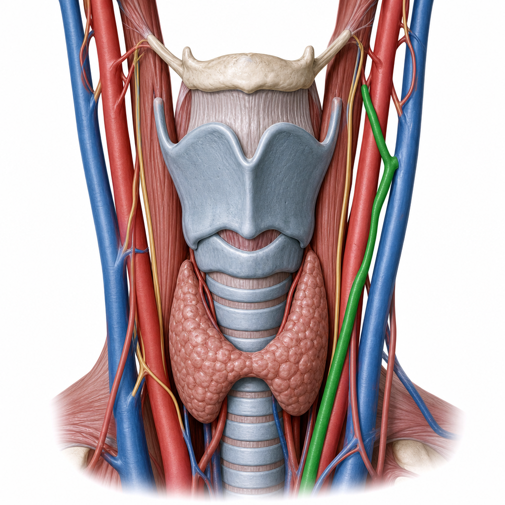
In the below diagram, the artery marked in complete green originates from the common carotid artery at the level of what anatomical structure?
Which of the following muscles is not affected by paralysis of the recurrent laryngeal nerve?
Chasaignac's tubercle is located at which anatomical site?
Which laryngeal cartilage is elastic?
Which muscle divides the neck into anterior and posterior triangles?
The lower border of the pharynx is the level of:
Killian's dehiscence is seen in:
Practice by Chapter
Cervical Fascia
Practice Questions
Triangles of the Neck
Practice Questions
Deep Structures of the Neck
Practice Questions
Thyroid and Parathyroid Glands
Practice Questions
Vasculature of the Neck
Practice Questions
Lymphatic Drainage
Practice Questions
Cervical Plexus
Practice Questions
Root of the Neck
Practice Questions
Applied Anatomy and Clinical Correlations
Practice Questions
Surface Anatomy of the Neck
Practice Questions
Want unlimited practice?
Get full access to all questions, explanations, and performance tracking.
Scan to download app