The submandibular gland is divided into superficial and deep parts by which muscle?
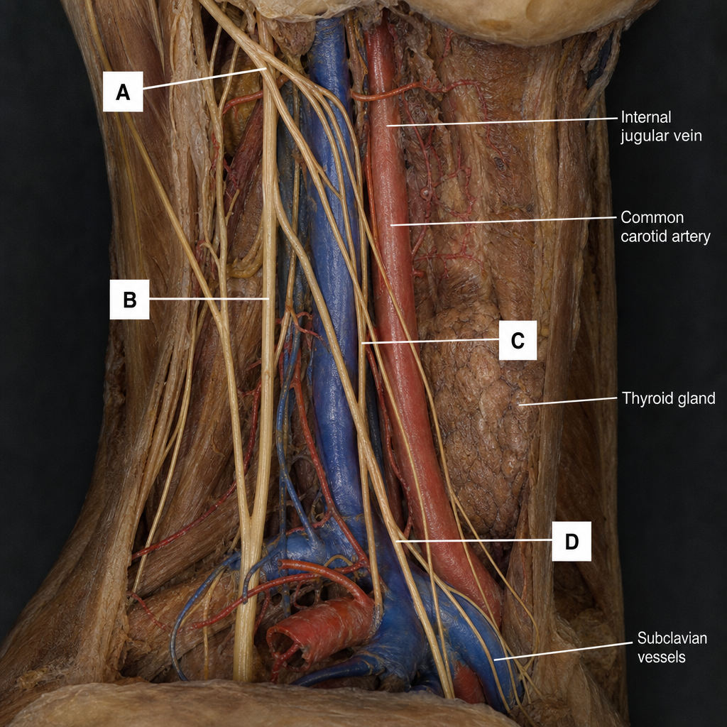
Which of the following is an extrinsic membrane of the larynx?
Which of the following statements about the larynx is false?
Which muscles are tensors of the vocal cords?
Which of the following hyoid muscles is an important landmark in both the anterior and posterior triangles of the neck?
Following a surgical procedure on the right side of the neck, a patient cannot raise their right arm above the horizontal position and cannot shrug their right shoulder. Which of the following nerves was injured?
Retroauricular incision in children less than 2 years old may cause damage to which cranial nerve?
Identify the nerve indicated by the arrow, which is important during surgery of the overlying organ.

The inferior thyroid artery arises from which of the following vessels?
The sinus of Morgagni is present in which of the following anatomical regions?
Explanation: ### Explanation **Correct Answer: C. Mylohyoid** The **mylohyoid muscle** is known as the "diaphragm of the mouth." It acts as the key anatomical landmark that divides the submandibular gland into a large superficial part and a small deep part. The gland is "U-shaped," wrapping around the posterior free border of the mylohyoid muscle. * The **superficial part** lies in the submandibular triangle (below the muscle). * The **deep part** lies in the floor of the mouth (above the muscle), extending forward to the sublingual gland. **Analysis of Incorrect Options:** * **A. Digastric:** The two bellies of the digastric muscle (along with the mandible) form the boundaries of the submandibular triangle where the gland is located, but they do not divide the gland itself. * **B. Geniohyoid:** This muscle lies superior to the mylohyoid and forms part of the floor of the mouth. It is medial to the deep part of the submandibular gland but does not divide it. * **D. Stylohyoid:** This muscle passes superficial to the carotid arteries and attaches to the hyoid bone; it relates to the posterior pole of the gland but has no role in its structural division. **High-Yield Clinical Pearls for NEET-PG:** * **Wharton’s Duct:** The submandibular duct emerges from the **deep part** of the gland and runs forward on the hyoglossus muscle. * **Nerve Relations:** The **lingual nerve** loops under Wharton’s duct (from lateral to medial) – "The nerve loops under the duct." * **Bimanual Palpation:** Because the gland wraps around the mylohyoid, it is best examined by placing one finger inside the mouth and the other under the jaw. * **Secretions:** The submandibular gland is a **mixed** gland (predominantly serous) and is the most common site for salivary stones (**Sialolithiasis**) due to its upward ductal course and viscous secretions.
Explanation: ### Explanation The membranes and ligaments of the larynx are classified into two groups: **Extrinsic** and **Intrinsic**. **1. Why the Thyrohyoid Membrane is Correct:** Extrinsic membranes connect the laryngeal cartilages to structures outside the larynx (such as the hyoid bone or the trachea). The **Thyrohyoid membrane** connects the upper border of the thyroid cartilage to the hyoid bone. It is a fibroelastic membrane pierced by the **internal laryngeal nerve** and the **superior laryngeal artery**, a high-yield anatomical landmark. **2. Why the Other Options are Incorrect:** Intrinsic membranes connect the laryngeal cartilages to each other, forming the internal framework of the larynx. * **Quadrangular Membrane (A):** An intrinsic membrane extending between the epiglottis and arytenoid cartilages. Its free lower margin forms the **vestibular ligament** (false vocal cord). * **Cricothyroid Membrane (C):** An intrinsic membrane connecting the cricoid and thyroid cartilages. * **Conus Elasticus (D):** Also known as the cricovocal membrane, this is the lower part of the intrinsic fibroelastic membrane. Its free upper margin forms the **vocal ligament** (true vocal cord). **Clinical Pearls for NEET-PG:** * **Cricothyroidotomy:** Performed through the median cricothyroid ligament (part of the intrinsic membrane) for emergency airway access. * **Internal laryngeal Nerve:** Injury during thyroid surgery (while piercing the thyrohyoid membrane) leads to loss of sensation above the vocal cords, increasing the risk of aspiration. The recurrent laryngeal nerve relations are also critical during such surgeries [1]. * **Vocal Cord Structure:** Remember that the **Vocalis muscle** and **Vocal ligament** (from conus elasticus) form the true vocal folds.
Explanation: ### Explanation **Why Option C is the correct answer (The False Statement):** The nerve supply of the laryngeal muscles follows a specific rule that is frequently tested. The **Recurrent Laryngeal Nerve (RLN)** supplies all the intrinsic muscles of the larynx [2] **except** for the **Cricothyroid** muscle. The Cricothyroid is instead supplied by the **External Laryngeal Nerve** (a branch of the Superior Laryngeal Nerve). Option C incorrectly swaps this relationship. **Analysis of Other Options:** * **Option A (True):** The laryngeal skeleton consists of 9 cartilages. There are **3 unpaired** (Thyroid, Cricoid, Epiglottis) and **3 paired** (Arytenoid, Corniculate, Cuneiform). * **Option B (True):** In adults, the larynx extends vertically from the level of the **C3 to the C6** vertebrae. In children, it is positioned higher (around C2-C3). * **Option D (True):** The **Cricothyroid** muscle tilts the thyroid cartilage forward, lengthening and tensing the vocal folds. It is known as the "singer’s muscle." **High-Yield Clinical Pearls for NEET-PG:** 1. **Safety Muscle:** The **Posterior Cricoarytenoid** is the only abductor of the vocal cords. Paralysis of this muscle leads to airway obstruction. 2. **Nerve Injury:** * Injury to the **External Laryngeal Nerve** (often during thyroidectomy) results in a weak, husky voice due to the inability to tense the vocal cords. * Unilateral **RLN injury** causes hoarseness; bilateral injury can cause stridor and respiratory distress [1]. 3. **Sensory Supply:** Above the vocal folds, sensation is carried by the **Internal Laryngeal Nerve**; below the folds, it is carried by the **Recurrent Laryngeal Nerve** [2].
Explanation: The vocal cords' tension and length are regulated by the intrinsic muscles of the larynx to control the pitch of the voice. ### **Explanation of the Correct Answer** The correct answer is **D (Cricothyroid and internal thyroarytenoid)** because these two muscles act as the primary tensors, though they do so in different ways: 1. **Cricothyroid:** This is the **primary tensor**. It tilts the thyroid cartilage forward or the cricoid cartilage backward, increasing the distance between the thyroid and arytenoid cartilages. This elongates and tenses the vocal cords, raising the pitch. 2. **Internal Thyroarytenoid (Vocalis):** This muscle lies within the vocal fold. By contracting locally, it adjusts the internal tension of the folds, allowing for fine-tuning of pitch and tone. ### **Analysis of Incorrect Options** * **Posterior cricoarytenoid (Option A):** This is the **sole abductor** of the vocal cords (the "safety muscle of the larynx"). It opens the glottis for breathing but does not act as a tensor. * **Lateral cricoarytenoid (Option B):** This is a primary **adductor**, closing the glottis by rotating the arytenoid cartilages medially. * **Interarytenoid (Options A, B, C):** These muscles (transverse and oblique) act as **adductors** by pulling the two arytenoid cartilages together, closing the posterior part of the rima glottidis. * **Thyroarytenoid (External part):** Generally acts as a **relaxor** of the vocal cords by pulling the arytenoids toward the thyroid cartilage, shortening the folds. ### **NEET-PG High-Yield Pearls** * **Nerve Supply:** All intrinsic muscles of the larynx are supplied by the **Recurrent Laryngeal Nerve (RLN)**, **EXCEPT** the **Cricothyroid**, which is supplied by the **External Laryngeal Nerve**. * **Safety Muscle:** The Posterior Cricoarytenoid is the only muscle that opens the airway; bilateral paralysis leads to respiratory distress. * **Vocalis:** Often considered the "fine-tuner" of the vocal cords, it is technically the medial-most fibers of the thyroarytenoid muscle.
Explanation: ### Explanation The **Omohyoid muscle** is a unique infrahyoid muscle consisting of two bellies (superior and inferior) connected by an intermediate tendon. It serves as a critical anatomical landmark because its bellies traverse both major triangles of the neck: 1. **Anterior Triangle:** The **superior belly** of the omohyoid travels vertically to divide the anterior triangle into the **carotid triangle** (above) and the **muscular triangle** (below). 2. **Posterior Triangle:** The **inferior belly** crosses the lower part of the posterior triangle, dividing it into the larger **occipital triangle** (above) and the smaller **supraclavicular (subclavian) triangle** (below). #### Analysis of Incorrect Options: * **Geniohyoid & Mylohyoid (A & B):** These are suprahyoid muscles located in the floor of the mouth. They contribute to the boundaries of the **submental triangle** but do not extend into the posterior triangle. * **Sternohyoid (D):** This is the most superficial infrahyoid muscle. While it is a key component of the **muscular triangle** in the anterior neck, it does not cross into the posterior triangle. #### NEET-PG High-Yield Pearls: * **Intermediate Tendon:** The intermediate tendon of the omohyoid is held down to the clavicle by a process of the **deep cervical fascia (pretracheal layer)**. * **Nerve Supply:** Like most infrahyoid muscles (except thyrohyoid), the omohyoid is supplied by the **Ansa Cervicalis (C1-C3)**. * **Surgical Landmark:** The omohyoid is a key landmark during **neck dissections**; the internal jugular vein (IJV) lies deep to the point where the omohyoid crosses the carotid sheath.
Explanation: **Explanation:** The patient’s clinical presentation—inability to shrug the shoulder and inability to abduct the arm above the horizontal (90 degrees)—points directly to a paralysis of the **Trapezius muscle**. 1. **Why the Spinal Accessory Nerve (CN XI) is correct:** The Spinal Accessory nerve provides motor innervation to the Sternocleidomastoid and the Trapezius. The Trapezius is essential for shrugging the shoulders and, more importantly, for rotating the scapula upward. This upward rotation is required to abduct the arm beyond 90 degrees (overhead). Because the nerve has a superficial course in the **posterior triangle of the neck**, it is highly vulnerable to injury during surgical procedures like lymph node biopsies or radical neck dissections. 2. **Why the other options are incorrect:** * **Axillary nerve:** Innervates the Deltoid. Injury would cause loss of abduction up to 90 degrees, but it does not affect shrugging or overhead rotation. * **Great auricular nerve:** A sensory branch of the cervical plexus (C2, C3). Injury would cause numbness over the parotid gland and lower ear, not motor deficits. * **Greater occipital nerve:** A sensory nerve (posterior ramus of C2) supplying the scalp. It has no motor function related to the shoulder. **High-Yield NEET-PG Pearls:** * **Surface Anatomy:** The Spinal Accessory nerve enters the posterior triangle at the junction of the upper 1/3 and middle 1/3 of the Sternocleidomastoid and exits at the junction of the middle 1/3 and lower 1/3 of the Trapezius. * **The "Drooping Shoulder":** A classic sign of CN XI injury is a drooping shoulder on the affected side due to Trapezius atrophy. * **Scapular Winging:** While the Long Thoracic nerve causes "true" winging (medial border), CN XI injury causes "lateral" winging of the scapula.
Explanation: The correct answer is **VII (Facial Nerve)**. **Why it is correct:** In infants and children under 2 years of age, the **mastoid process** is not yet developed (it begins developing around the end of the first year and matures by age 2). Consequently, the **stylomastoid foramen**, through which the Facial Nerve (CN VII) exits the skull, is located very superficially and laterally on the skull base. In adults, the mastoid process provides a bony "shield" that protects the nerve. In young children, a retroauricular (behind the ear) incision can easily transect the nerve because it lies just beneath the skin and fascia, lacking this bony protection. **Why the other options are incorrect:** * **A (VIII - Vestibulocochlear):** This nerve remains deep within the internal acoustic meatus and the petrous part of the temporal bone. It is not at risk during superficial skin incisions. * **C (VI - Abducens):** This nerve has a long intracranial course and exits via the superior orbital fissure to supply the lateral rectus muscle. It is nowhere near the retroauricular region. * **D (V - Trigeminal):** While the Trigeminal nerve provides sensory innervation to the face, the specific risk in this surgical approach is the motor trunk of the Facial nerve. The Great Auricular nerve (C2, C3) provides sensation to this area, not CN V. **High-Yield NEET-PG Pearls:** * **Developmental Milestone:** The mastoid process develops due to the pull of the **Sternocleidomastoid muscle** as the child begins to hold their head up and move. * **Surgical Safety:** To avoid CN VII injury in infants, incisions should be placed more superiorly or further away from the anticipated exit point of the stylomastoid foramen. * **Other superficial structures:** The **Parotid gland** is also more superiorly placed in neonates, further exposing the facial nerve branches.
Explanation: ***Recurrent laryngeal nerve*** - Runs in the **tracheoesophageal groove** closely related to the **thyroid gland**, making it vulnerable during thyroidectomy and requiring careful identification to prevent injury. - Injury causes **vocal cord paralysis** leading to **hoarseness** and potential **airway compromise**, making it the most critical nerve during thyroid surgery. *Superior laryngeal nerve* - Divides into **internal** and **external branches** above the level of the thyroid gland, not running in the tracheoesophageal groove. - The **internal branch** provides **sensory innervation** to the supraglottis, while the **external branch** innervates the **cricothyroid muscle**. *External laryngeal nerve* - A **branch of the superior laryngeal nerve** that innervates the **cricothyroid muscle** for **vocal cord tension**. - Located more **superiorly** and **laterally** compared to the recurrent laryngeal nerve, not in the tracheoesophageal groove. *Internal laryngeal nerve* - The **sensory branch** of the superior laryngeal nerve providing sensation to the **larynx above the vocal cords**. - Pierces the **thyrohyoid membrane** and does not have the same close anatomical relationship to the thyroid gland as the recurrent laryngeal nerve.
Explanation: **Explanation:** The **inferior thyroid artery** is the primary vessel supplying the posterior and inferior aspects of the thyroid gland. It is a major branch of the **thyrocervical trunk**, which itself arises from the first part of the **subclavian artery** [1]. This anatomical relationship is crucial for understanding the blood supply to the neck and the parathyroid glands. **Analysis of Options:** * **Thyrocervical trunk (Correct):** This trunk gives off four main branches: the inferior thyroid, suprascapular, transverse cervical, and ascending cervical arteries [1]. * **Brachiocephalic trunk:** This vessel gives rise to the right common carotid and right subclavian arteries but does not directly give off the inferior thyroid artery. * **Internal carotid artery:** This artery has no branches in the neck; it enters the skull to supply the brain and eyes. * **External carotid artery:** This vessel gives rise to the **superior thyroid artery** (its first anterior branch). Confusing the superior and inferior thyroid arteries is a common examination pitfall. **High-Yield Clinical Pearls for NEET-PG:** * **Recurrent Laryngeal Nerve (RLN):** The inferior thyroid artery has a critical surgical relationship with the RLN [2]. During thyroidectomy, the artery is usually ligated far from the gland to avoid damaging the nerve. * **Parathyroid Supply:** The inferior thyroid artery is the **sole** or primary blood supply to both the superior and inferior parathyroid glands [1]. * **Thyroid Ima Artery:** In ~3-10% of individuals, an accessory artery (Artery of Neubauer) arises directly from the brachiocephalic trunk or aortic arch to supply the thyroid isthmus.
Explanation: ### Explanation The **Sinus of Morgagni** (also known as the pharyngeal recess or hiatus) is a gap in the pharyngeal wall located in the **Nasopharynx**. #### Why Nasopharynx is Correct The muscular wall of the pharynx is not continuous. There is a distinct gap between the upper border of the **Superior Constrictor muscle** and the **Base of the Skull** (petrous part of the temporal bone). This gap is the Sinus of Morgagni. It is reinforced by the pharyngobasilar fascia and serves as a conduit for several important structures to enter the pharynx: 1. **Auditory (Eustachian) tube** 2. **Levator veli palatini muscle** 3. **Ascending palatine artery** #### Why Other Options are Incorrect * **Oropharynx:** This region lies between the soft palate and the upper border of the epiglottis. The gap between the superior and middle constrictors (which transmits the stylopharyngeus muscle and glossopharyngeal nerve) is located here, but it is not termed the Sinus of Morgagni. * **Hypopharynx / Laryngopharynx:** These terms are synonymous. This region extends from the epiglottis to the lower border of the cricoid cartilage. The gaps here (between middle/inferior constrictors and below the inferior constrictor) transmit the laryngeal nerves and vessels, not the structures associated with Morgagni. #### High-Yield Clinical Pearls for NEET-PG * **Passavant’s Ridge:** A mucosal ridge in the nasopharynx formed by the palatopharyngeal sphincter; it helps in closing the nasopharyngeal isthmus during swallowing. * **Killian’s Dehiscence:** A weak area in the lower pharynx (between thyropharyngeus and cricopharyngeus) which is the site for **Zenker’s Diverticulum**. * **Rosenmüller's Fossa:** A deep recess behind the tubal elevation in the nasopharynx; it is the most common site of origin for **Nasopharyngeal Carcinoma**.
Cervical Fascia
Practice Questions
Triangles of the Neck
Practice Questions
Deep Structures of the Neck
Practice Questions
Thyroid and Parathyroid Glands
Practice Questions
Vasculature of the Neck
Practice Questions
Lymphatic Drainage
Practice Questions
Cervical Plexus
Practice Questions
Root of the Neck
Practice Questions
Applied Anatomy and Clinical Correlations
Practice Questions
Surface Anatomy of the Neck
Practice Questions
Get full access to all questions, explanations, and performance tracking.
Start For Free