Joint involved in movement of head from left to right.
What is the primary tensor of the vocal cords?
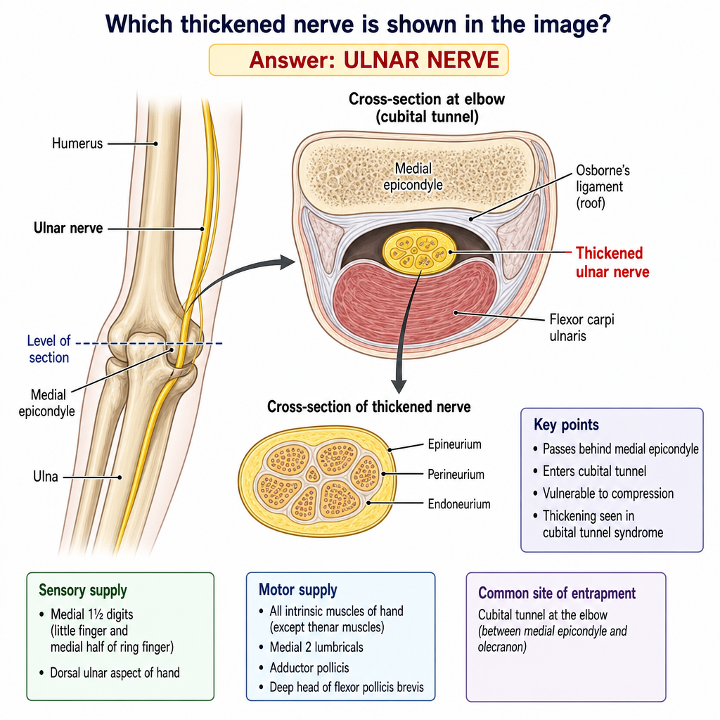
The main muscle affected in congenital muscular torticollis is?
Which of the following is not an extrinsic laryngeal membrane?
Inlet of larynx is formed by:
How are submandibular nodes classified?
Which muscle is the deepest in the anterior neck region?
What is Galen's anastomosis?
What is the nerve supply of the larynx above the level of the vocal cords?
Which of the following is NOT a content of the occipital triangle?
Explanation: ***Atlanto axial*** - The **atlantoaxial joint** is responsible for the **rotation of the head**, allowing for movements such as shaking the head "no." - This joint is a **pivot joint** formed between the atlas (C1 vertebra) and the axis (C2 vertebra), specifically the **dens** of the axis articulating with the anterior arch of the atlas. *Atlanto occipital* - The **atlanto-occipital joint** primarily facilitates **flexion and extension of the head** (nodding "yes" movement). - This joint connects the **atlas (C1)** to the **occipital bone** of the skull. *C2- C3 Joint* - The **C2-C3 joint** is a typical **intervertebral joint** in the cervical spine. - While it contributes to overall cervical spine mobility, it does not primarily mediate the **rotational movement** of the head. *C3- C4 Joint* - The **C3-C4 joint** is another **intervertebral joint** in the cervical spine. - Its main roles include some degree of **flexion, extension, and lateral bending**, but it is not the primary joint for head rotation.
Explanation: ***Cricothyroid*** - The **cricothyroid muscle** is the principal tensor of the vocal cords, responsible for stretching and thinning them. - It achieves this by tilting the **thyroid cartilage** forward relative to the **cricoid cartilage**, increasing the distance between the **thyroid** and **arytenoid cartilages**. *Lateral Cricoarytenoid* - This muscle primarily functions as an **adductor** of the vocal cords, bringing them together. - It rotates the **arytenoid cartilages** medially, closing the **rima glottidis**. *Thyroarytenoids* - The **thyroarytenoid muscles** are located within the vocal cords and primarily act to **shorten and relax** the vocal cords. - They also contribute to **adduction** and can internally tense the vocal folds, but their main role is *not* primary tension. *Posterior cricoarytenoids* - The **posterior cricoarytenoid muscles** are the *only* muscles responsible for **abducting** (opening) the vocal cords. - They rotate the **arytenoid cartilages** laterally, thus widening the **rima glottidis**.
Explanation: ***Sternocleidomastoid*** - Congenital muscular torticollis (CMT) is primarily caused by **unilateral fibrosis and shortening of the sternocleidomastoid muscle (SCM)**. - This leads to the characteristic **head tilt towards the affected side** and **chin rotation to the opposite side**. *Trapezius* - The trapezius muscle is primarily involved in **shrugging the shoulders**, extending and rotating the head and neck, but is not the main muscle affected in CMT. - While it can become secondarily tight in response to persistent head positioning, it is **not the primary pathological muscle** in CMT. *Scalenus Anticus* - The scalenus anticus (anterior scalene muscle) is involved in **neck flexion and elevation of the first rib** during forced inspiration. - It plays a role in various neck pain syndromes and brachial plexus compression, but it is **not the defining muscle in congenital muscular torticollis**. *Omohyoid* - The omohyoid is a **strap muscle of the neck** that depresses the hyoid bone. - It has no primary involvement in the **pathophysiology or clinical presentation of congenital muscular torticollis**.
Explanation: **Quadrangular membrane** - The quadrangular membrane is an **intrinsic laryngeal membrane**, originating and inserting within the larynx itself, forming the false vocal cords and epiglottic folds. - It does not connect the larynx to external structures like the hyoid bone or trachea. *Hyoepiglottic ligament* - This is an **extrinsic laryngeal ligament** that connects the anterior surface of the epiglottis to the body of the hyoid bone. - It helps anchor the epiglottis to a structure outside the larynx. *Cricotracheal membrane* - The cricotracheal membrane is an **extrinsic laryngeal membrane** connecting the inferior border of the cricoid cartilage of the larynx to the first tracheal ring. - It forms the connection between the larynx and the trachea, an external structure. *Thyrohyoid membrane* - This is an **extrinsic laryngeal membrane** that connects the superior border of the thyroid cartilage of the larynx to the first tracheal ring. - It provides a broad connection between the larynx and the hyoid bone, an external laryngeal structure.
Explanation: ***Aryepiglottic fold*** - The **inlet of the larynx** is the opening into the laryngeal cavity from the pharynx. - It is bordered anteriorly by the **epiglottis**, laterally by the **aryepiglottic folds**, and posteriorly by the **arytenoid cartilages** and **interarytenoid notch**. *False vocal cord* - The **false vocal cords** (ventricular folds) are located within the laryngeal cavity, inferior to the inlet. - They play a protective role but do not form the boundaries of the laryngeal inlet itself. *Folds from the base of the tongue to the epiglottis* - These folds, including the **glossoepiglottic folds**, connect the tongue to the epiglottis. - They are superior to the laryngeal inlet and are part of the oropharynx, not direct borders of the inlet. *Vocal cord* - The **true vocal cords** are responsible for voice production and are located deeper within the larynx, inferior to the false vocal cords. - They do not form any part of the laryngeal inlet.
Explanation: ***Level I neck nodes*** - Submandibular nodes, along with the **submental nodes**, are anatomically grouped as **Level I** in the regional neck lymph node classification [1]. - This classification is crucial for **staging head and neck cancers** and guiding surgical management [1], [3]. *Level II neck nodes* - Level II nodes are located in the **upper jugular region**, extending from the skull base to the caudal border of the hyoid bone, often including nodes around the **spinal accessory nerve**. - This level is distinct from the submandibular region and drains different anatomical areas. *Level III neck nodes* - Level III nodes are found in the **mid-jugular region**, from the caudal border of the hyoid bone to the caudal border of the cricoid cartilage, associated with the **internal jugular vein**. [2] - They primarily drain the oropharynx, hypopharynx, and larynx, not the submandibular region. *Level IV neck nodes* - Level IV nodes are located in the **lower jugular region**, from the caudal border of the cricoid cartilage to the clavicle, also associated with the **internal jugular vein** [2]. - These nodes drain the lower hypopharynx, larynx, and apical lung, and are anatomically distant from the submandibular area.
Explanation: ***Longus colli*** - The **longus colli** muscle is the **deepest muscle** located in the anterior neck region, running along the front of the cervical vertebral column from C1 to T3. - It lies in the **prevertebral layer**, deep to all other anterior neck structures including the carotid sheath, visceral compartment, and superficial muscles. - Its position directly anterior to the vertebral bodies makes it the deepest anterior neck muscle. *Platysma* - The platysma is the **most superficial muscle** of the neck, located just beneath the skin in the superficial fascia. - It is not a deep muscle and lies superficial to all other neck muscles. *Sternocleidomastoid* - The sternocleidomastoid is enclosed within the **investing layer of deep cervical fascia**, making it relatively superficial. - While prominent in the anterior and lateral neck, it is not the deepest anterior neck muscle. *Trapezius* - The trapezius is a large, **superficial muscle of the back and posterior neck**. - It is not located in the anterior neck and is a superficial, not deep, muscle.
Explanation: ***Internal laryngeal nerve and recurrent laryngeal nerve*** - **Galen's anastomosis** (also known as **ansa Galeni**) is the connection between the **internal laryngeal nerve** (a sensory branch of the superior laryngeal nerve) and the **recurrent laryngeal nerve** (motor branch of the vagus nerve). - This anastomosis occurs in the **laryngeal mucosa** and allows for communication between these two important nerves. - The **internal laryngeal nerve** provides **sensory innervation** to the larynx above the vocal cords, while the **recurrent laryngeal nerve** provides **motor innervation** to most intrinsic laryngeal muscles (except cricothyroid). - This anastomosis is clinically significant in understanding the complex innervation patterns of the larynx. *Recurrent laryngeal nerve and external laryngeal nerve* - While both nerves are branches of the vagus nerve and innervate laryngeal structures, this connection does not constitute **Galen's anastomosis**. - The recurrent laryngeal nerve innervates intrinsic laryngeal muscles (except cricothyroid), while the external laryngeal nerve innervates the **cricothyroid muscle** and is motor in nature. *None of the options* - This option is incorrect because the correct definition of **Galen's anastomosis** is provided in the first option. *Recurrent laryngeal nerve and sympathetic trunk* - While there may be sympathetic contributions to laryngeal innervation, this does not represent **Galen's anastomosis**. - The sympathetic trunk provides autonomic innervation but the specific named anastomosis refers to the connection between the internal laryngeal and recurrent laryngeal nerves.
Explanation: ***Superior laryngeal*** - The **superior laryngeal nerve** branches into the internal and external laryngeal nerves. The **internal laryngeal nerve** (a branch of the superior laryngeal nerve) provides all sensory innervation to the larynx **above the vocal cords**. - It also carries **parasympathetic fibers** to the laryngeal glands in this region. *Recurrent laryngeal* - The **recurrent laryngeal nerve** provides sensory innervation to the larynx **below the vocal cords** [1]. - It also innervates all of the intrinsic muscles of the larynx except for the cricothyroid muscle [1]. *Glossopharyngeal* - The **glossopharyngeal nerve (CN IX)** primarily provides sensory innervation to the **posterior one-third of the tongue**, tonsils, pharynx, and middle ear. - It does not directly provide sensory innervation to the larynx. *External laryngeal nerve* - The **external laryngeal nerve**, a branch of the superior laryngeal nerve, is primarily **motor** and innervates the **cricothyroid muscle**. - It provides **no sensory innervation** to any part of the larynx.
Explanation: Suprascapular nerve - The **suprascapular nerve** originates from the brachial plexus and supplies the supraspinatus and infraspinatus muscles; it travels through the suprascapular notch and is not found within the occipital triangle. - Its primary course and innervation are associated with the shoulder, entirely separate from the neck region defining the occipital triangle. *Great auricular nerve* - The **great auricular nerve** emerges from the cervical plexus and supplies sensory innervation to the skin over the parotid gland, mastoid process, and auricle, courses superficially across the sternocleidomastoid in the region of the occipital triangle. - It is a recognized content of the posterior triangle of the neck, which encompasses the occipital triangle. *Lesser occipital nerve* - The **lesser occipital nerve** arises from the cervical plexus at C2 and C3, providing sensory innervation to the skin of the neck and scalp posterior to the auricle. - It ascends along the posterior border of the sternocleidomastoid muscle, placing it within the boundaries of the occipital triangle. *Occipital artery* - The **occipital artery** is a branch of the external carotid artery that supplies blood to the posterior scalp. - It traverses the apex of the posterior triangle (including the occipital triangle) as it ascends to the back of the head.
Cervical Fascia
Practice Questions
Triangles of the Neck
Practice Questions
Deep Structures of the Neck
Practice Questions
Thyroid and Parathyroid Glands
Practice Questions
Vasculature of the Neck
Practice Questions
Lymphatic Drainage
Practice Questions
Cervical Plexus
Practice Questions
Root of the Neck
Practice Questions
Applied Anatomy and Clinical Correlations
Practice Questions
Surface Anatomy of the Neck
Practice Questions
Get full access to all questions, explanations, and performance tracking.
Start For Free