
Neck — MCQs

On this page
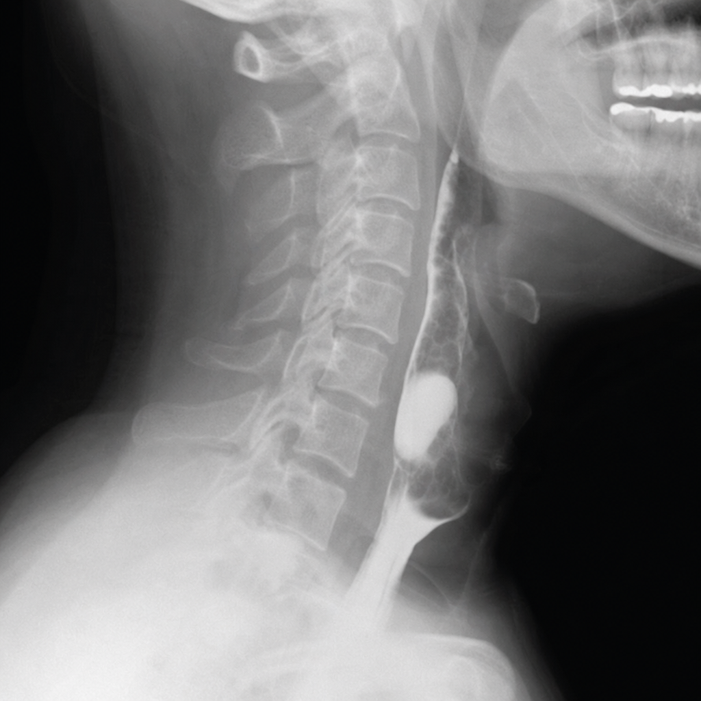
A 58-year-old male patient presents with halitosis, mild dysphagia and regurgitation of previous day's food. On radiological examination this is the presentation of the patient. What is the location of this presentation? (Recent NEET Pattern 2016-17)
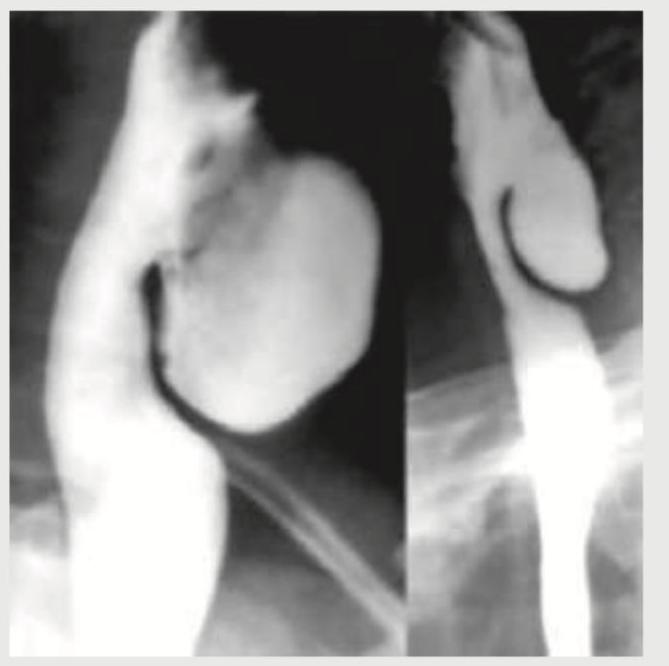
A 58-year-old male patient presents with halitosis, mild dysphagia and regurgitation of previous day food. Barium study is performed. Where is the location of this presentation?
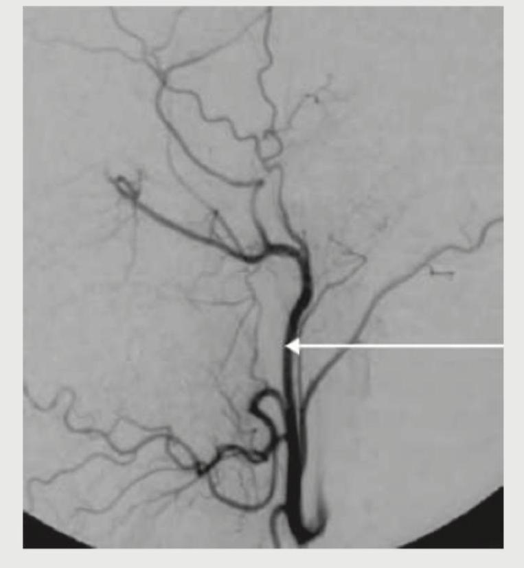
What is the name of the marked blood vessel, which is a branch of external carotid artery?
Important landmark in submandibular gland dissection is:
Third part of vertebral artery is related to which of the following ?
Inferior thyroid artery supplies which of the following structures? 1. Thyroid 2. Parathyroid 3. Esophagus 4. Thymus
During a surgical procedure involving the posterior triangle of the neck, which of the following muscles forms its anterior boundary?
During a thyroidectomy, a surgeon must carefully identify and preserve the parathyroid glands. These glands are most commonly located posterior to which part of the thyroid gland?
A 45-year-old male patient presents with difficulty swallowing and hoarseness that has progressively worsened over the past month. During physical examination, the physician notices that the patient's left vocal cord is paralyzed. The paralysis is most likely due to compression of which of the following nerves?
Adam's apple in males is formed by the
Practice by Chapter
Cervical Fascia
Practice Questions
Triangles of the Neck
Practice Questions
Deep Structures of the Neck
Practice Questions
Thyroid and Parathyroid Glands
Practice Questions
Vasculature of the Neck
Practice Questions
Lymphatic Drainage
Practice Questions
Cervical Plexus
Practice Questions
Root of the Neck
Practice Questions
Applied Anatomy and Clinical Correlations
Practice Questions
Surface Anatomy of the Neck
Practice Questions
Want unlimited practice?
Get full access to all questions, explanations, and performance tracking.
Scan to download app